

Overview
Chordoma and chondrosarcoma can both involve the clivus, the central bone at the base of the skull, but they differ in their biology, pathology, and treatment planning. Local control depends on the anatomy, how much tumor can be removed safely, the pathology, and the radiation strategy.
For these reasons, care emphasizes expert pathology and imaging review and long-term surveillance rather than a single-operation narrative. Treatment is individualized and coordinated across surgery and radiation oncology. This page is educational and does not replace specialist evaluation.
What this evaluation should clarify
A focused evaluation is meant to answer a few key questions:
- What objective evidence distinguishes chordoma and chondrosarcoma from each other and from look-alike conditions?
- Which anatomic, neurologic, or tumor-related contributors must be considered?
- Which combination of surgery, radiation, and surveillance best fits the pathology, the findings, and your goals?
Living with chordoma and chondrosarcoma? The next step is a quiet, unhurried conversation.
Evaluation and treatment pathway
Care usually follows a stepwise pathway:
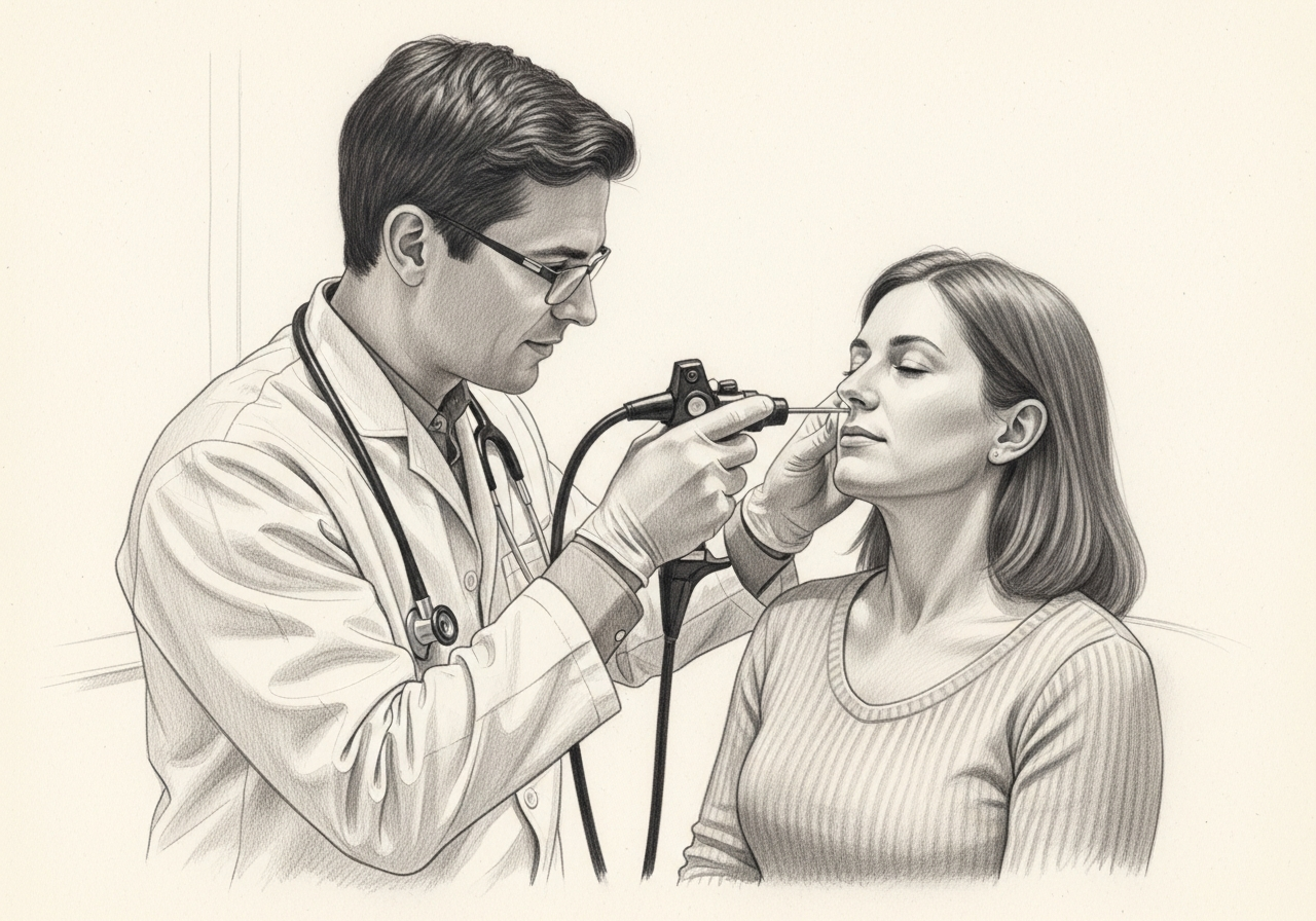
- Review the symptom pattern, duration, triggers, prior treatment, operations, medications, and relevant medical history.
- Use high-resolution MRI and CT to define the bone and soft-tissue extent, with expert pathology to distinguish these tumors from one another and from related lesions. Prior surgery, radiation, and molecular findings may change planning.
- Identify important look-alikes, complications, and contributors before settling on a diagnosis.
- Treatment often combines maximal safe removal with specialized radiation, tailored to the pathology, any residual disease, prior treatment, and the risk to function. Long-term imaging surveillance is important because recurrence can occur.
- Set a clear follow-up plan, including symptom goals, objective reassessment, and imaging or surveillance when appropriate.
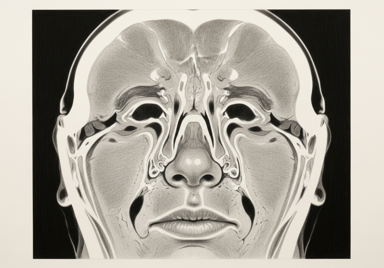
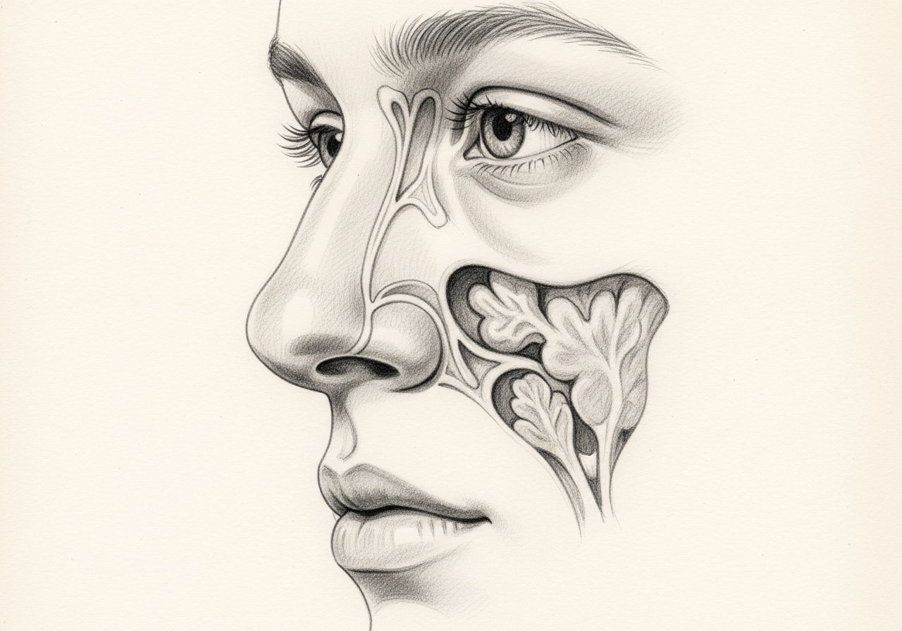
Clival anatomy and tumor differences
Chordomas and chondrosarcomas arise at the base of the skull, often in the clivus, a central bone in front of the brainstem. Chordomas develop from remnants of the embryonic notochord, while chondrosarcomas arise from cartilage-forming cells, often slightly off the midline. Both sit near critical nerves and vessels, which shapes how they are treated.
Endoscopic, open, and combined surgery
Surgery aims to remove as much tumor as safely possible while protecting vital structures. Depending on the location and extent, this may be done endoscopically through the nose, through an open approach, or with a combination. Complete removal is not always achievable because of the surrounding anatomy, which is one reason radiation is frequently part of treatment.
Radiation planning
Radiation therapy is often used after surgery to treat residual or microscopic tumor, frequently with specialized high-precision techniques that aim to spare nearby critical structures. The plan is individualized and made with radiation oncology.
Long-term surveillance and recurrence
Because chordomas and chondrosarcomas can recur, sometimes years later, long-term imaging surveillance is essential. Ongoing follow-up allows recurrence to be detected and addressed as early as possible.
What to bring to your consultation
To make the most of your visit, bring or securely share the records that can change the plan:
- Imaging files and reports, such as CT or MRI
- Endoscopy or operative findings
- Pathology results
- Laboratory results
- Notes from prior treatment
- A current medication list
- The specific question you would like answered
When to seek urgent care
New double vision, vision loss, facial numbness, difficulty swallowing, weakness, severe headache, or other rapidly progressive neurologic symptoms requires urgent evaluation. An online form or routine appointment request is not an emergency service; for these symptoms, seek immediate in-person care.
Medical review
This page is a patient-education resource reviewed by the responsible Norelle Health clinician before publication. It does not replace an in-person evaluation. If symptoms are severe or rapidly worsening, seek immediate medical care.
Specialists who treat chordoma and chondrosarcoma
Dr. Adrian Ong
MD
Board-Certified Facial Plastic & Reconstructive and Head & Neck Surgeon
Dr. Adrian Ong is a board-certified surgeon who practices exclusively on the face, head, and neck, with expertise spanning rhinoplasty, sinus surgery, facial trauma, reconstruction, and sleep surgery.
- Functional and aesthetic rhinoplasty (including revision)
- Sinus surgery and complex revision sinus surgery
- Facial trauma and nasal fractures
- Head and neck cancer surgery and microvascular reconstruction
Also caring for this area

Not sure who to see? Our patient coordination team can help match you with the right specialist.
(212) 444-8006Frequently Asked Questions
Chordoma is a rare tumor that arises from remnants of the embryonic notochord, while chondrosarcoma is a cartilage-forming tumor. Both can occur at the skull base near cranial nerves and major blood vessels.
High-resolution MRI and CT define the bone and soft-tissue extent, and expert pathology distinguishes these tumors from one another and from related lesions. Prior surgery, radiation, and molecular findings can change planning.
Treatment often combines maximal safe removal with specialized radiation, tailored to the pathology, any residual disease, prior treatment, and the risk to function. Long-term imaging surveillance is important because these tumors can recur.
New double vision, vision loss, facial numbness, difficulty swallowing, weakness, severe headache, or other rapidly progressive neurologic symptoms requires urgent evaluation.
Clinical References
These independent resources from medical and professional organizations offer further reading. They are provided for general education and do not replace a consultation with a clinician.
Related Procedures
1 of 2 · Endoscopic Skull Base Surgery
Related Conditions
Request a consultation for chordoma and chondrosarcoma
Schedule an evaluation with our team to review your symptoms and the appropriate next steps.