
Overview
Craniopharyngiomas arise near the pituitary gland, the optic pathways, and the hypothalamus. Although they are not cancers, their location means treatment decisions must balance tumor control with hormone function, vision, cognition, appetite, and long-term quality of life.
For this reason, care is approached as a multidisciplinary decision rather than a single surgical procedure. Planning draws on neurosurgery, endocrinology, ophthalmology, and, when an endonasal route is appropriate, skull-base rhinology, with long-term follow-up after treatment. This page is educational and does not replace specialist evaluation.
What this evaluation should clarify
A focused evaluation is meant to answer a few key questions:
- What objective evidence distinguishes a craniopharyngioma from conditions that can look similar?
- Which structures are involved, including the optic pathways, pituitary, and hypothalamus, and which other contributors must be considered?
- Which path, from observation to surgery, radiation, or a combination, best fits the findings and your goals?
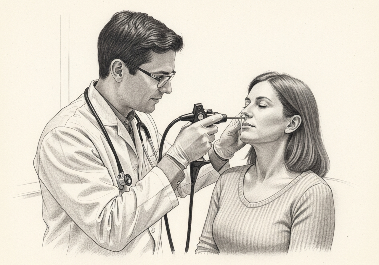
Living with craniopharyngioma? The next step is a quiet, unhurried conversation.
Evaluation and treatment pathway
Care usually follows a stepwise pathway:
- Review the symptom pattern, duration, triggers, prior treatment, operations, medications, and relevant medical history.
- Use MRI and CT to define the cystic, solid, and calcified parts of the tumor and their relationship to the optic pathways, pituitary stalk, hypothalamus, and vessels. A hormonal (endocrine) evaluation and formal vision testing are central to planning.
- Identify important look-alikes, complications, and contributors before settling on a diagnosis.
- Options may include observation for selected cases, surgery, cyst-directed treatment, radiation, or combinations. The extent of removal is weighed against the risk to function and the chance of recurrence, which calls for long-term imaging, endocrine, and visual follow-up.
- Set a clear follow-up plan, including symptom goals, objective reassessment, medication response, and imaging or surveillance when appropriate.
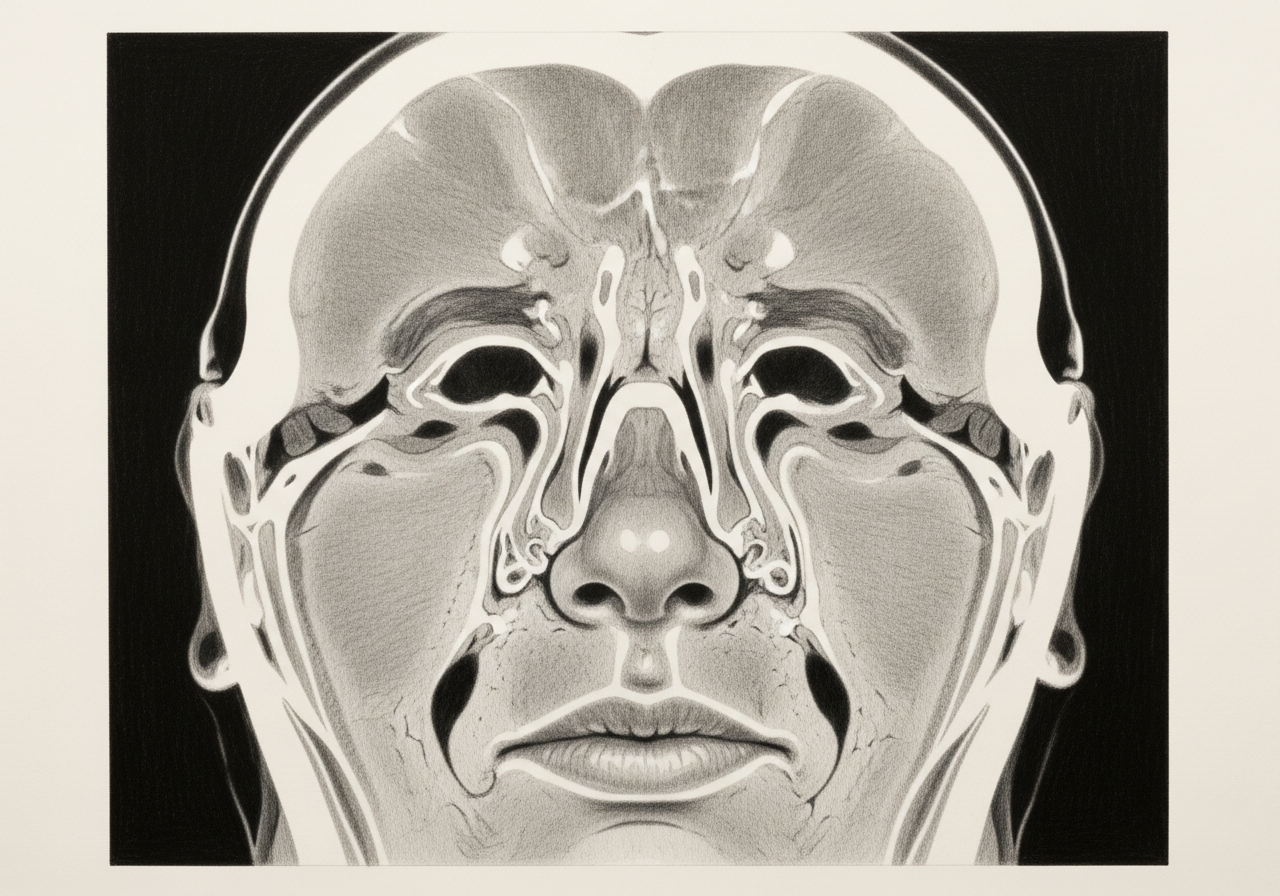
Vision, hormonal, and hypothalamic symptoms
Because of its location, a craniopharyngioma can compress the optic pathways and cause vision changes, affect the pituitary and lead to hormonal symptoms such as excessive thirst, fatigue, or growth changes, and involve the hypothalamus, which can affect appetite, weight, and temperature regulation. Headache is also common.
Observation, surgery, and radiation
Management is individualized. Some situations are observed, while others are treated with surgery, radiation, or a combination. The plan depends on the size and location of the tumor, the symptoms it is causing, and the patient's overall health, and it is made by a multidisciplinary team.
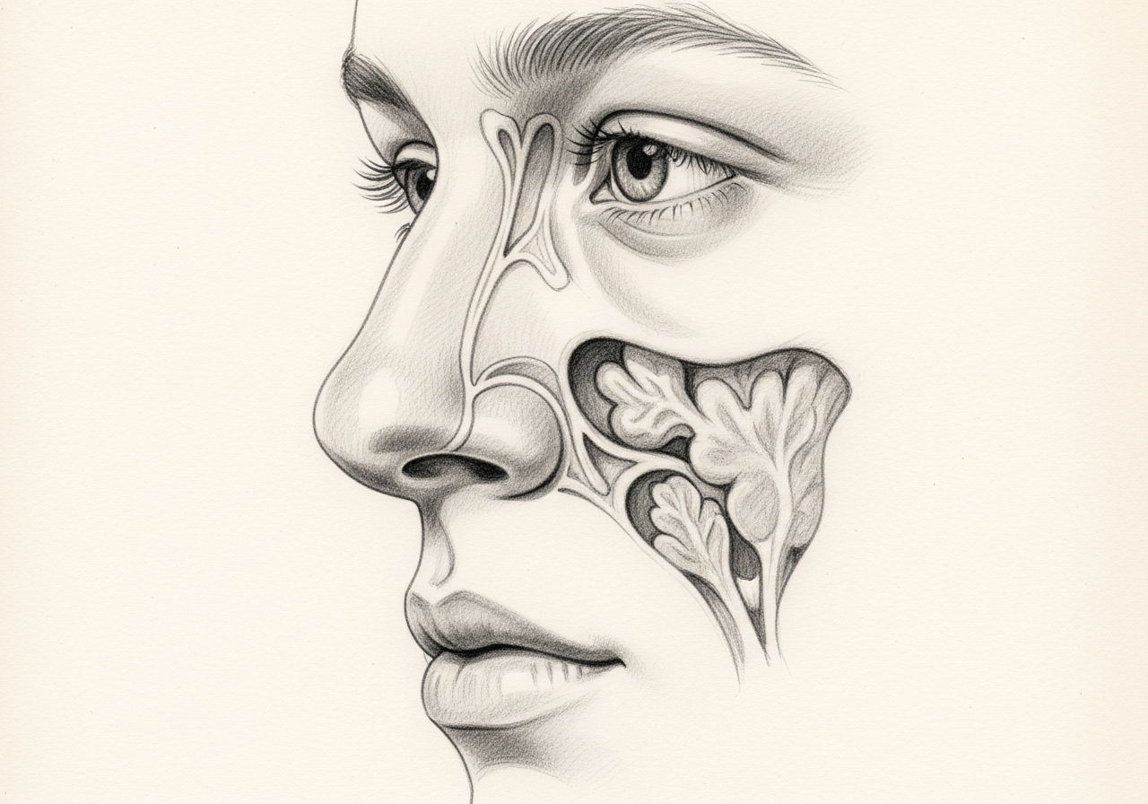
Endoscopic versus open routes
When surgery is chosen, some craniopharyngiomas can be approached endoscopically through the nose (an endonasal route), while others require an open approach. The route depends on the tumor's exact location, its extension, and its relationship to nearby nerves and vessels. The goal is to relieve pressure and obtain tissue while protecting vital structures.
Long-term endocrine and visual follow-up
Because the tumor and its treatment can affect hormones and vision, long-term follow-up with endocrinology and vision testing is important. Hormone replacement is managed by endocrinology when needed, and imaging is used to monitor for recurrence.

What to bring to your consultation
To make the most of your visit, bring or securely share the records that can change the plan:
- Imaging files and reports, such as CT or MRI
- Endoscopy or operative findings
- Pathology results
- Laboratory results
- Notes from prior treatment
- A current medication list
- The specific question you would like answered
When to seek urgent care
Sudden vision change, severe headache, vomiting, confusion, marked weakness, seizure, or acute hormonal instability requires emergency assessment. An online form or routine appointment request is not an emergency service; for these symptoms, seek immediate in-person care.
Medical review
This page is a patient-education resource reviewed by the responsible Norelle ENT clinician before publication. It does not replace an in-person evaluation. If symptoms are severe or rapidly worsening, seek immediate medical care.
Specialists who treat craniopharyngioma

Dr. Adrian Ong
MD
Board-Certified Facial Plastic & Reconstructive and Head & Neck Surgeon
Dr. Adrian Ong is a board-certified surgeon who practices exclusively on the face, head, and neck, with expertise spanning rhinoplasty, sinus surgery, facial trauma, reconstruction, and sleep surgery.
- Functional and aesthetic rhinoplasty (including revision)
- Sinus surgery and complex revision sinus surgery
- Facial trauma and nasal fractures
- Head and neck cancer surgery and microvascular reconstruction
Also caring for this area


Not sure who to see? Our patient coordination team can help match you with the right specialist.
(212) 444-8006Frequently Asked Questions
A craniopharyngioma is a rare, usually non-spreading tumor near the pituitary stalk and hypothalamus. Because of its location, it can affect vision, hormone function, and nearby brain structures.
MRI and CT define the cystic, solid, and calcified parts of the tumor and their relationship to the optic pathways, pituitary stalk, hypothalamus, and blood vessels. A hormonal (endocrine) evaluation and formal vision testing are central to planning.
Not always. Options may include observation for selected cases, surgery, cyst-directed treatment, radiation, or combinations. The extent of removal is weighed against the risk to function and the chance of recurrence, with long-term imaging, endocrine, and visual follow-up.
Sudden vision change, severe headache, vomiting, confusion, marked weakness, seizure, or acute hormonal instability requires emergency assessment.
Clinical References
These independent resources from medical and professional organizations offer further reading. They are provided for general education and do not replace a consultation with a clinician.
Related Procedures

1 of 3 · Endoscopic Pituitary Surgery

Request a consultation for craniopharyngioma
Schedule an evaluation with our team to review your symptoms and the appropriate next steps.