

Overview
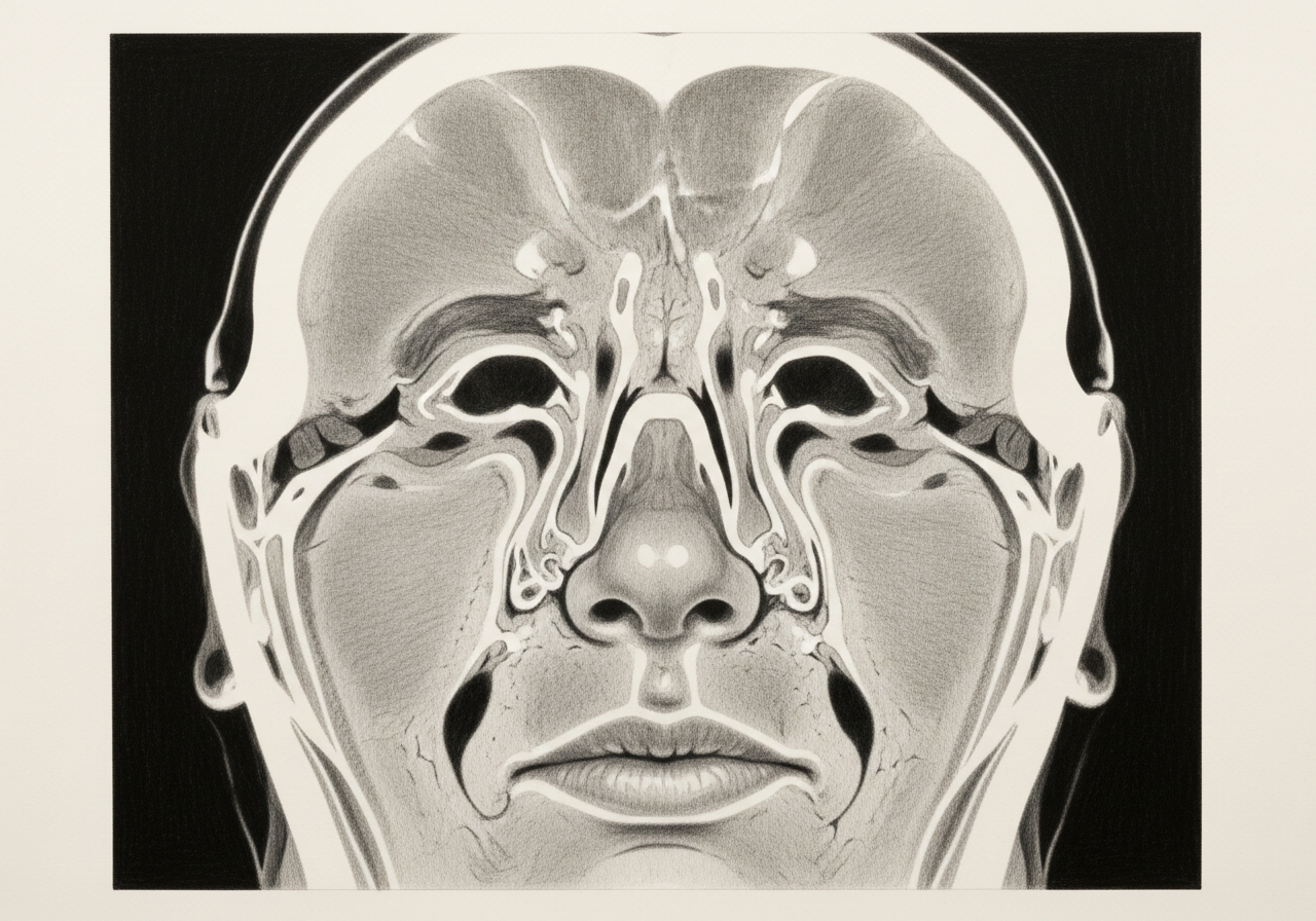
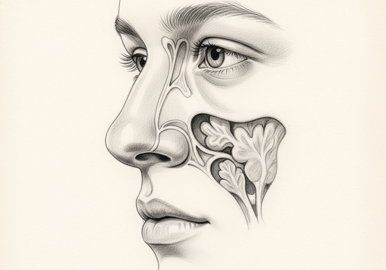
A meningocele or encephalocele is more than a nasal mass. Tissue from inside the skull, or its coverings, protrudes through a defect in the skull base and may be associated with a cerebrospinal fluid (CSF) leak, an increased risk of infection, or elevated pressure of the fluid around the brain.
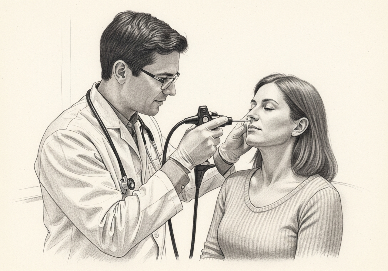
Because the lesion can be continuous with the space around the brain, it should not be biopsied like a routine nasal polyp. Imaging is used first to define the defect and what it contains, so that evaluation and any repair can be planned safely by a team with ENT and neurosurgical expertise.
What this evaluation should clarify
A focused evaluation is meant to answer a few key questions:
- What objective evidence distinguishes a meningocele or encephalocele from conditions that can look similar, such as a nasal polyp or other mass?
- Which anatomic, neurologic, infectious, or pressure-related contributors need to be considered?
- Which medical, procedural, surgical, or multidisciplinary path best fits the findings and your goals?
Living with meningocele and encephalocele? The next step is a quiet, unhurried conversation.
Evaluation and treatment pathway
Care usually follows a stepwise pathway:
- Review the symptom pattern, duration, triggers, prior treatment, operations, medications, and relevant medical history.
- Use CT to define the bony defect and MRI to characterize the soft-tissue contents and their relationship to structures inside the skull. The evaluation also looks for a CSF leak, prior trauma or surgery, congenital factors, and signs of elevated pressure of the fluid around the brain.
- Identify important look-alikes, complications, and contributors before settling on a diagnosis.
- Treatment depends on symptoms, whether there is a leak, the location, the herniated tissue, infection risk, and patient factors. Endoscopic repair is used for many anterior skull-base defects, while selected lesions are observed or require a different surgical approach.
- Set a clear follow-up plan, including symptom goals, objective reassessment, and imaging or surveillance when appropriate.
Relationship to CSF rhinorrhea
Because these defects create an opening in the skull base, they are often associated with cerebrospinal fluid (CSF) leaking into the nose, known as CSF rhinorrhea. A persistent clear, watery nasal drip, especially from one side, can be a sign and should be evaluated.
Congenital, traumatic, and spontaneous causes
Encephaloceles and meningoceles may be present from birth, result from trauma or prior surgery, or develop spontaneously. Spontaneous cases are sometimes associated with elevated pressure of the fluid around the brain, which is an important factor in planning treatment.
Endoscopic versus open repair
Many defects in this region can be repaired endoscopically through the nose, avoiding an external incision, by carefully sealing the defect and reinforcing the skull base. Open repair is reserved for selected larger or more complex defects. Repair is performed by a team with ENT and neurosurgical expertise.
Pressure evaluation and recurrence
When elevated pressure of the fluid around the brain contributes to the defect, addressing that pressure is important to reduce the chance of recurrence after repair. Follow-up confirms the repair remains durable over time.
What to bring to your consultation
To make the most of your visit, bring or securely share the records that can change the plan:
- Imaging files and reports, such as CT or MRI
- Endoscopy or operative findings
- Pathology results
- Laboratory results
- Notes from prior treatment
- A current medication list
- The specific question you would like answered
When to seek urgent care
Fever, neck stiffness, severe headache, confusion, seizure, new neurologic symptoms, persistent clear nasal drainage, or symptoms after head trauma require urgent or emergency assessment. An online form or routine appointment request is not an emergency service; for these symptoms, seek immediate in-person care.
Medical review
This page is a patient-education resource reviewed by the responsible Norelle Health clinician before publication. It does not replace an in-person evaluation. If symptoms are severe or rapidly worsening, seek immediate medical care.
Specialists who treat meningocele and encephalocele

Dr. Adrian Ong
MD
Board-Certified Facial Plastic & Reconstructive and Head & Neck Surgeon
Dr. Adrian Ong is a board-certified surgeon who practices exclusively on the face, head, and neck, with expertise spanning rhinoplasty, sinus surgery, facial trauma, reconstruction, and sleep surgery.
- Functional and aesthetic rhinoplasty (including revision)
- Sinus surgery and complex revision sinus surgery
- Facial trauma and nasal fractures
- Head and neck cancer surgery and microvascular reconstruction
Also caring for this area

Not sure who to see? Our patient coordination team can help match you with the right specialist.
(212) 444-8006Frequently Asked Questions
A meningocele is a herniation of the meninges, the coverings of the brain, through a skull-base defect. An encephalocele also contains brain tissue. Either can be continuous with the nose or sinuses.
CT defines the bony defect, and MRI characterizes the soft-tissue contents and their relationship to structures inside the skull. The evaluation also looks for a CSF leak, prior trauma or surgery, congenital factors, and signs of elevated pressure of the fluid around the brain.
Treatment depends on symptoms, whether there is a leak, the location, the herniated tissue, infection risk, and patient factors. Endoscopic repair is used for many anterior skull-base defects, while selected lesions are observed or require a different surgical approach.
Fever, neck stiffness, severe headache, confusion, seizure, new neurologic symptoms, persistent clear drainage, or symptoms after head trauma require urgent or emergency assessment.
Clinical References
These independent resources from medical and professional organizations offer further reading. They are provided for general education and do not replace a consultation with a clinician.
Related Procedures

1 of 3 · Endoscopic CSF Leak Repair

Request a consultation for meningocele and encephalocele
Schedule an evaluation with our team to review your symptoms and the appropriate next steps.