

About the Procedure
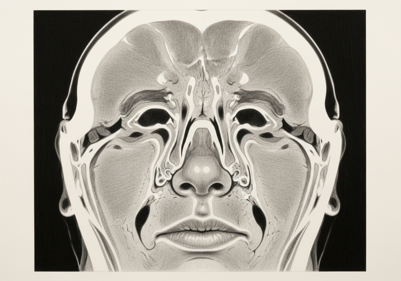
The skull base is the floor of the brain and the roof of the nose and sinuses. Skull-base reconstruction rebuilds the barrier between the cranial cavity and the nose after a cerebrospinal fluid (CSF) leak is repaired or a tumor is removed, so that CSF stays where it belongs and the brain is protected from infection.
The closure is matched to the size and location of the defect, how much CSF flow is expected, any prior radiation or surgery, the tissue available, and the patient's healing and pressure risks. Options range from small free grafts for low-flow defects to multilayer vascularized flap reconstruction for larger, high-flow defects.
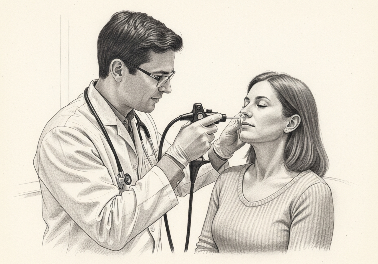
At Norelle Health, skull-base reconstruction is performed endoscopically through the nose in most cases, in coordination with neurosurgery when the brain or its coverings are involved. The goal is a durable, watertight repair with the least disruption to nasal function that the situation allows.
What this evaluation should clarify
This page is meant to help you understand a few key decisions about skull-base reconstruction:
- Whether the underlying diagnosis and the plan to treat the defect or leak are established with the right imaging and testing
- How the choice of closure compares across options, from a small graft to a vascularized flap or a combined approach
- Which factors — defect size and location, CSF flow, prior radiation or surgery, and your healing and pressure risks — change the recommendation
Considering skull base reconstruction? The next step is a quiet, unhurried conversation.
Evaluation and treatment pathway
- Confirm the diagnosis, the goal of treatment, and the reasons a less invasive approach is not enough.
- Planning begins with the expected or existing defect, the tissue available, any prior operations or radiation, infection status, CSF pressure, and the route used to treat the underlying problem.
- Options range from small free grafts to multilayer vascularized flap reconstruction or open and combined approaches. The least extensive closure that reliably fits the defect is chosen, rather than using one technique for every case.
- After the lesion or leak is addressed, reconstructive materials are positioned to separate the nose from the cranial cavity. A nasoseptal flap or other vascularized tissue may be used for larger or higher-flow defects, with donor-site and nasal effects discussed beforehand.
- Postoperative care protects the closure, watches for leakage and meningitis, and manages the nasal donor site. Activity, straining, saline care, packing, and follow-up are individualized.
Goals of reconstruction
The main goals of skull base reconstruction are to create a watertight seal that keeps CSF inside the space around the brain, to restore the barrier that protects against infection such as meningitis, and to support healing of the surgical area.
A secondary goal is to preserve nasal function as much as possible. The plan balances a secure repair against the effects of borrowing tissue from the nose, choosing the least disruptive option likely to provide a durable seal.
Low-flow versus high-flow defects
Reconstruction is tailored to how much CSF is expected to pass through the defect. A low-flow defect, such as a small leak, may be sealed with grafts alone. A high-flow defect, such as a large opening into the fluid-filled spaces after major tumor or pituitary surgery, usually needs a more robust, often vascularized repair.
Classifying the defect this way helps the team choose materials and techniques that match the risk, rather than treating every defect the same way.
Free grafts, local flaps and vascularized nasoseptal flap
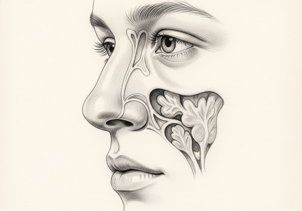
Several reconstructive options are used, often in layers. Free grafts use tissue such as the lining or a small amount of cartilage or bone placed over the defect. Local flaps and a vascularized nasoseptal flap, a piece of the septal lining that keeps its own blood supply, can be rotated to cover larger or higher-flow defects.
The nasoseptal flap is widely used for large skull-base repairs because its blood supply supports reliable healing. The choice of technique depends on the defect, prior surgery, and available tissue.
Lumbar drainage and sealants only when appropriate
In some higher-risk repairs, a temporary lumbar drain may be used for a short period to lower CSF pressure and protect the reconstruction while it heals. It is not needed for every case and is reserved for selected situations.
Tissue sealants and supporting materials may be added to reinforce the repair. These are adjuncts to a well-constructed reconstruction, not substitutes for it, and are used selectively based on the defect and surgeon judgment.
Donor-site and nasal effects
Because reconstruction often borrows tissue from the nose, there can be nasal effects during healing, including crusting, congestion, and temporary changes in smell. When a nasoseptal flap is used, the area it came from heals with crusting over several weeks and needs nasal care.
These effects are weighed against the benefit of a secure repair. The team aims to use the tissue needed for a durable seal while limiting the impact on nasal function.
What to bring to your consultation
Bringing the right records helps make the most of your visit. Where possible, bring or securely transfer:
- Imaging files and reports (such as CT or MRI of the sinuses and skull base)
- Any prior endoscopy or operative findings
- Pathology results, if a biopsy or surgery has been done
- Relevant laboratory results
- Notes from prior treatment and a current medication list
- The specific question you would most like answered
For complex skull-base care, your records may be reviewed with the specialists, including neurosurgery, involved in that part of your evaluation.
When to seek urgent care
Some symptoms need urgent or emergency assessment rather than a routine appointment. Seek care for constant clear, watery nasal drainage, a fever with a stiff neck, a severe headache, a change in vision, confusion, a seizure, major bleeding, or rapidly worsening illness after reconstruction.
These can signal a leak, meningitis, or another time-sensitive problem. An online form or routine appointment request is not an emergency service.
Medical review
This page is a patient-education resource reviewed by the responsible Norelle Health clinician before publication. It does not replace an in-person evaluation. Constant clear, watery nasal drainage, high fever, or severe headache with a stiff neck after skull-base surgery can signal a leak or infection and should prompt immediate medical care.
Specialists who perform skull base reconstruction
Dr. Adrian Ong
MD
Board-Certified Facial Plastic & Reconstructive and Head & Neck Surgeon
Dr. Adrian Ong is a board-certified surgeon who practices exclusively on the face, head, and neck, with expertise spanning rhinoplasty, sinus surgery, facial trauma, reconstruction, and sleep surgery.
- Functional and aesthetic rhinoplasty (including revision)
- Sinus surgery and complex revision sinus surgery
- Facial trauma and nasal fractures
- Head and neck cancer surgery and microvascular reconstruction
Also caring for this area

Not sure who to see? Our patient coordination team can help match you with the right specialist.
(212) 444-8006Frequently Asked Questions
Planning begins with the expected or existing defect, the tissue available, any prior operations or radiation, infection status, CSF pressure, and the route used to treat the underlying problem.
After the lesion or leak is addressed, reconstructive materials are positioned to separate the nose from the cranial cavity. A nasoseptal flap or other vascularized tissue may be used for larger or higher-flow defects, with donor-site and nasal effects discussed beforehand.
Options range from small free grafts to multilayer vascularized flap reconstruction or open and combined approaches. The least extensive closure that reliably fits the defect is chosen, rather than using one technique for every case.
Postoperative care protects the closure, watches for leakage and meningitis, and manages the nasal donor site. Activity, straining, saline care, packing, and follow-up are individualized.
Clinical References
These independent resources from medical and professional organizations offer further reading. They are provided for general education and do not replace a consultation with a clinician.
Related Conditions

Related Procedures

1 of 3 · Endoscopic CSF Leak Repair
Request a consultation about skull base reconstruction
Schedule a consultation with our team to discuss whether this procedure is the right option for you.