
Overview
The frontal sinus drains through a narrow, highly variable pathway close to the orbit and skull base. Persistent disease, scarring, mucoceles, or prior surgery call for precise imaging review and a plan tailored to the anatomy rather than a one-size-fits-all approach to sinus surgery. Forehead pain alone is not a reliable sign of frontal disease, so evaluation relies on nasal endoscopy, CT imaging, and a review of any prior surgery before deciding between medical therapy and surgical options.
What this evaluation should clarify
A focused evaluation is designed to answer a few key questions:
- What objective evidence distinguishes frontal sinus disease from look-alike conditions?
- Which anatomic, inflammatory, dental, neurologic, infectious, or tumor-related factors may be contributing?
- Which medical, procedural, surgical, or multidisciplinary path best fits the findings and your goals?
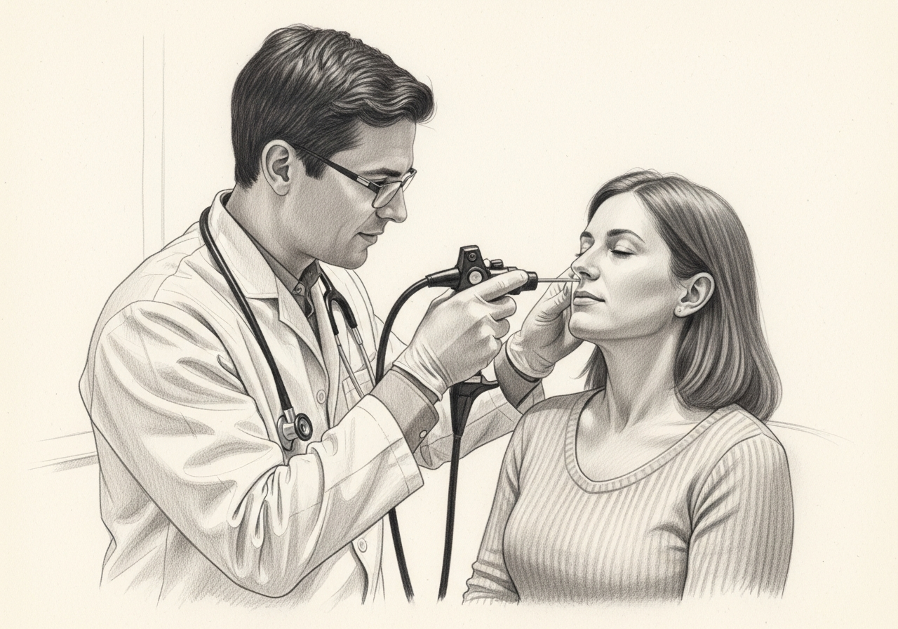
Living with frontal sinus disease? The next step is a quiet, unhurried conversation.
Evaluation and treatment pathway
Care generally follows a stepwise path:
- Clarify the symptom pattern, duration, triggers, prior treatment, operations, medications, and relevant medical history.
- Use a focused history, nasal endoscopy, and thin-cut CT review to define the frontal recess, prior surgical changes, cells that narrow drainage, and any extension toward the orbit or skull base.
- Identify important look-alikes, complications, and contributors before settling on a definitive diagnosis.
- Consider medical therapy for inflammatory disease, and reserve surgery for persistent objective disease, complications, mucoceles, tumors, or failed prior treatment, matching the extent to the anatomy and disease rather than automatically escalating.
- Set a measurable follow-up plan covering symptom goals, objective reassessment, medication response, and imaging or surveillance when appropriate.
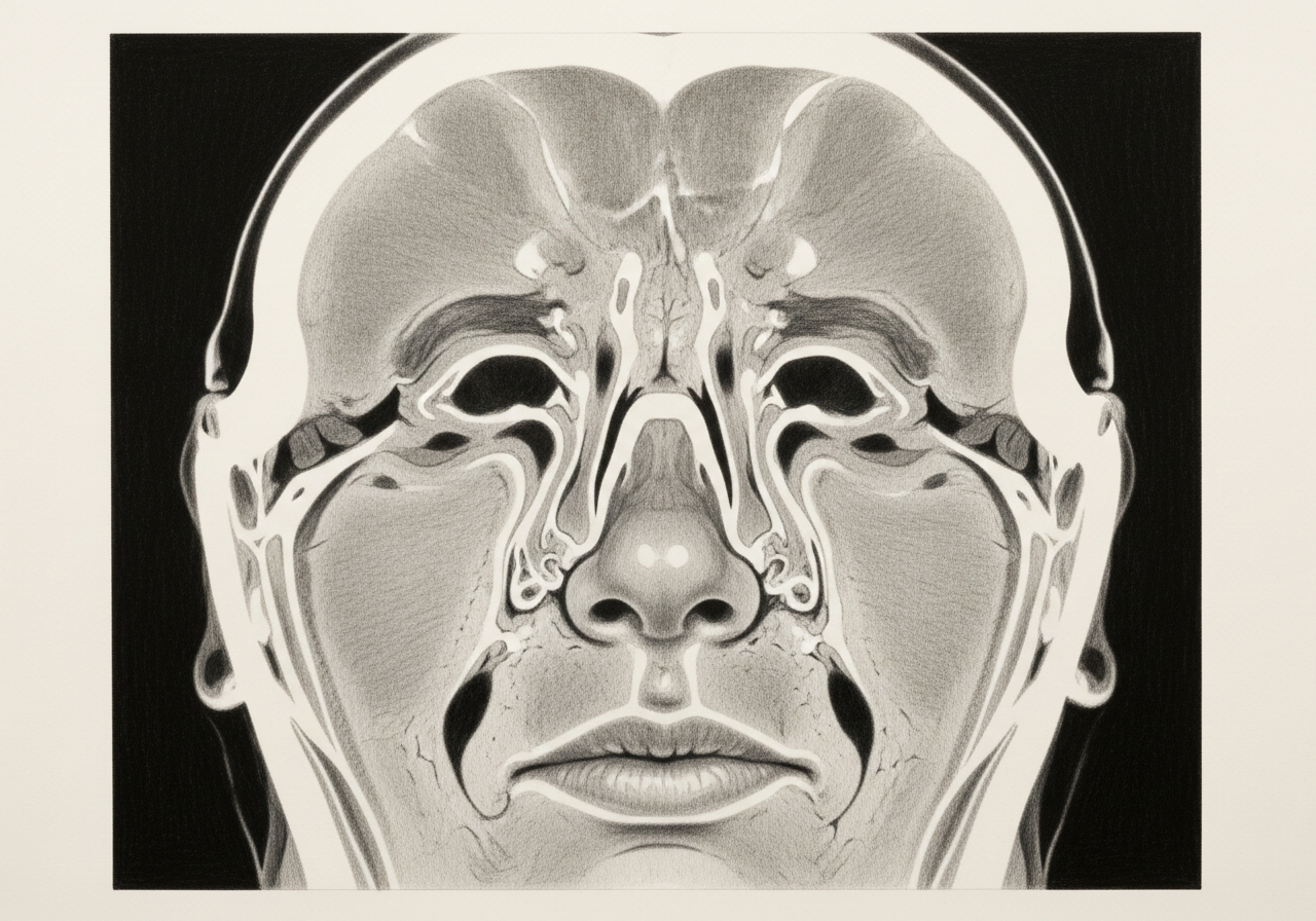
Frontal drainage anatomy
The frontal sinuses drain through a narrow, curving channel often called the frontal recess. The anatomy varies from person to person and can include small cells that crowd the pathway. Because the channel is tight, even modest inflammation or scarring can block drainage and lead to recurring disease.

Symptoms and why forehead pain alone is not diagnostic
Frontal disease can cause forehead pressure, congestion, and drainage, but forehead pain by itself is common and often has non-sinus causes such as tension or migraine. A diagnosis relies on examination and imaging rather than the location of pain alone.
Differential: inflammation, polyps, mucocele, and tumor
Frontal symptoms can come from inflammatory sinus disease, nasal polyps, a mucocele that traps and expands mucus, or, less commonly, a benign or malignant tumor. Distinguishing these guides whether medical therapy, surgery, or further work-up is appropriate.
Treatment options
Treatment is matched to the cause and severity:
- Medical therapy with saline irrigation, topical steroids, and treatment of infection
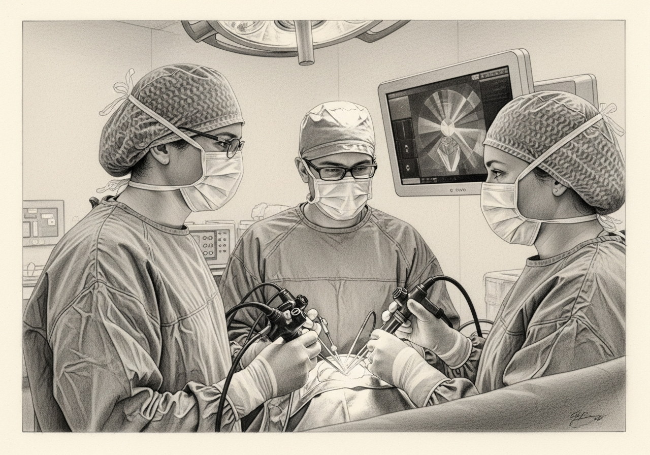
- Standard endoscopic frontal sinus surgery to open the drainage pathway
- Extended (Draf) procedures that enlarge the frontal outflow in selected complex or revision cases
- Open approaches in rare situations where endoscopic access is not sufficient
Not every patient needs surgery, and the least invasive approach that addresses the problem is generally preferred.
Scar prevention and follow-up
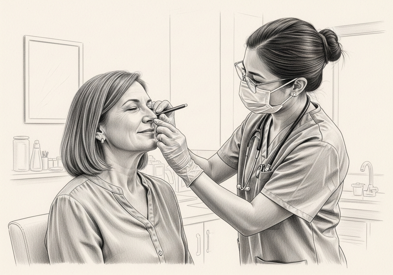
Because the frontal pathway is narrow, scarring and re-narrowing are the main reasons disease can return after surgery. Postoperative care, topical therapy, and follow-up endoscopy help keep the channel open and detect early narrowing.
What to bring to your consultation
Bringing the right records helps make the visit focused and useful. Where available, gather:
- Imaging files and reports, including CT and any MRI
- Endoscopy or operative findings from prior care
- Pathology results from any biopsy or surgery
- Relevant laboratory results
- Notes from prior treatment and a current medication list
- The specific question you would like answered
When to seek urgent care
New forehead swelling, eye swelling, vision change, severe frontal headache, fever with toxicity, neurologic symptoms, or rapidly worsening pain requires urgent evaluation.
Medical review
This page is a patient-education resource reviewed by the responsible Norelle ENT clinician before publication. It does not replace an in-person evaluation. If symptoms are severe or rapidly worsening, seek immediate medical care.
Specialists who treat frontal sinus disease
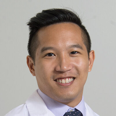
Dr. Adrian Ong
MD
Board-Certified Facial Plastic & Reconstructive and Head & Neck Surgeon
Dr. Adrian Ong is a board-certified surgeon who practices exclusively on the face, head, and neck, with expertise spanning rhinoplasty, sinus surgery, facial trauma, reconstruction, and sleep surgery.
- Functional and aesthetic rhinoplasty (including revision)
- Sinus surgery and complex revision sinus surgery
- Facial trauma and nasal fractures
- Head and neck cancer surgery and microvascular reconstruction
Also caring for this area


Not sure who to see? Our patient coordination team can help match you with the right specialist.
(212) 444-8006Frequently Asked Questions
Frontal sinus disease includes inflammatory blockage, infection, scarring, mucoceles, and other processes affecting the frontal sinus or its drainage pathway.
A focused history, nasal endoscopy, and thin-cut CT review define the frontal recess, prior surgical changes, cells that narrow drainage, and any extension toward the orbit or skull base.
Medical therapy may control inflammatory disease. Surgery is considered for persistent objective disease, complications, mucoceles, tumors, or failed prior treatment, with the extent matched to the anatomy and disease rather than automatically escalating.
New forehead swelling, eye swelling, vision change, severe frontal headache, fever with toxicity, neurologic symptoms, or rapidly worsening pain requires urgent evaluation.
Clinical References
These independent resources from medical and professional organizations offer further reading. They are provided for general education and do not replace a consultation with a clinician.
Related Procedures

1 of 3 · Advanced Frontal Sinus Surgery

Request a consultation for frontal sinus disease
Schedule an evaluation with our team to review your symptoms and the appropriate next steps.