

Overview
Salivary gland cancer is a group of uncommon cancers that arise in the glands that make saliva. The major salivary glands are the paired parotid, submandibular, and sublingual glands. Hundreds of smaller minor salivary glands are distributed through the lips, mouth, palate, throat, nose, and upper airway. A cancer can therefore present as a lump in front of the ear, beneath the jaw, inside the mouth, or in another head and neck site.
Unlike many cancers that are dominated by one cell type, salivary malignancies include a wide range of histologies with different growth patterns and treatment considerations. Some behave slowly over years; others are more aggressive or have a tendency to travel along nerves. The diagnosis must therefore be more specific than “a salivary tumor.” Expert pathology review, careful imaging, and a surgical plan that accounts for nearby nerves and blood vessels are central to treatment.
Surgery is the main treatment for many localized salivary gland cancers. Depending on the gland, tumor type, stage, and pathology, treatment may also include neck dissection, postoperative radiation, systemic therapy, or reconstruction. The aim is complete cancer treatment while preserving facial movement, swallowing, speech, shoulder function, and appearance whenever oncologically appropriate.
How we approach the decision
This page is meant to help you understand the choices that shape a salivary gland cancer plan, then connect each one to the next appropriate step. Three questions usually frame the discussion:
- Has an experienced pathologist confirmed the exact histology and grade? A parotid tumor and other salivary masses can look alike, so expert review matters.
- Can the facial nerve or other critical structures be preserved without compromising cancer removal during parotidectomy or salivary gland surgery?
- Does the plan need neck treatment, head and neck reconstruction, nerve repair, or postoperative radiation? For rare or previously treated disease, a head and neck second opinion can clarify the path.
Living with salivary gland cancer? The next step is a quiet, unhurried conversation.
Why this condition deserves a focused evaluation
Most salivary gland lumps are not cancer, yet a malignant tumor cannot be identified reliably by feel alone. Pain, rapid growth, skin fixation, numbness, or weakness of the face may raise concern, but some cancers remain painless and mobile. A focused evaluation is important because the biopsy method, imaging, and operative approach should be planned together rather than treating the mass as an isolated lump.
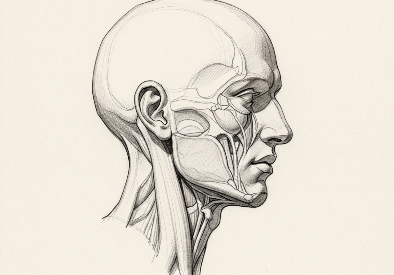
The anatomic stakes differ by gland. The facial nerve branches through the parotid gland and controls movement of the forehead, eyelids, cheek, and mouth. The submandibular gland sits near nerves that move the lower lip and tongue. Minor salivary tumors may arise beside structures needed for speech and swallowing. In addition, certain histologies can spread along nerves or to lymph nodes, lungs, bone, or other sites. An organized workup identifies the likely diagnosis, maps the relationship to critical structures, and allows the patient to understand the tradeoffs before surgery.
Anatomy and where the disease begins
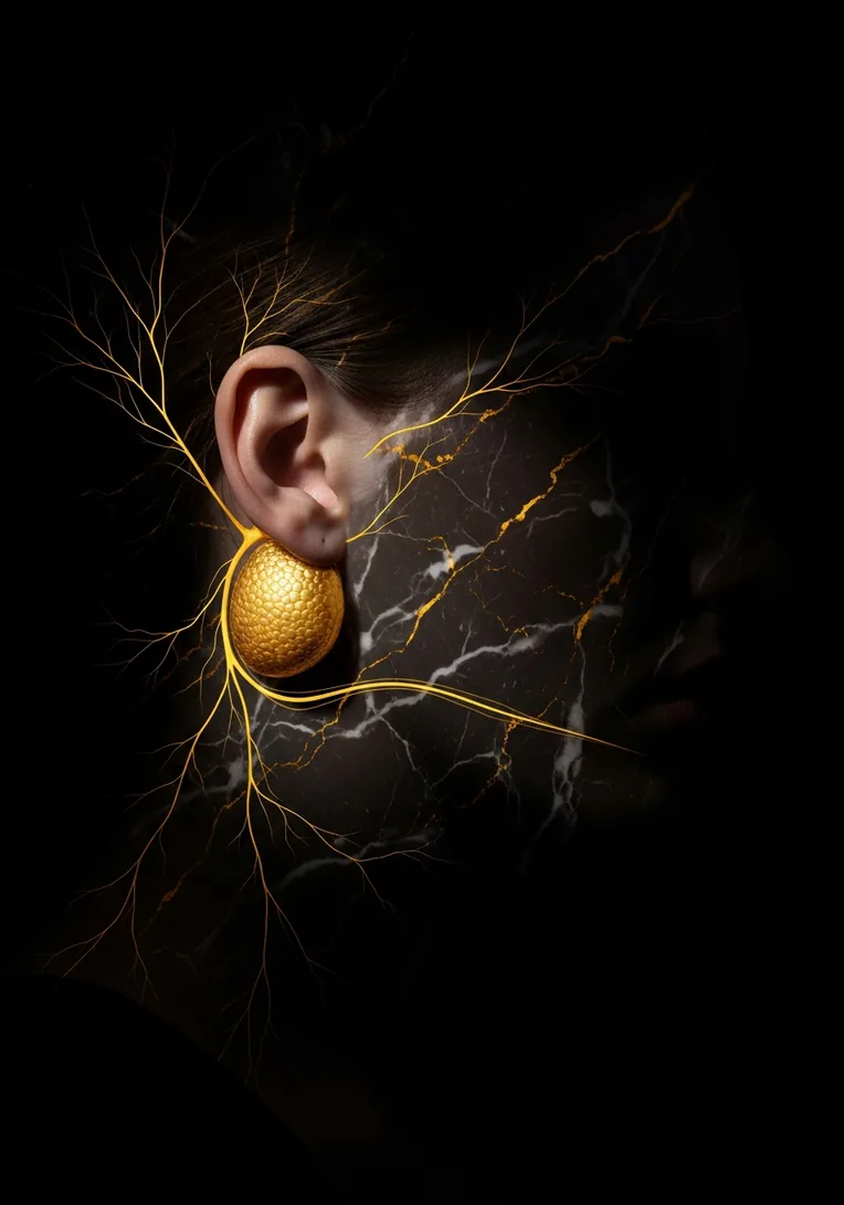
The parotid is the largest salivary gland and lies in front of and below the ear. The facial nerve enters the gland and divides into branches that travel between superficial and deep portions. A parotid tumor may arise outside the nerve, around it, or—less commonly—directly involve it. Deep-lobe tumors can extend toward the throat and may not be obvious from the outside.
The submandibular glands lie beneath the jaw. Nearby structures include the marginal mandibular branch of the facial nerve, the lingual nerve, the hypoglossal nerve, and the duct that carries saliva into the mouth. The sublingual glands sit beneath the tongue. Minor salivary glands are scattered through the palate, lips, cheek, tongue, nasal cavity, and pharynx.
Cancer behavior also depends on microscopic type and grade. Common malignant categories include mucoepidermoid carcinoma, adenoid cystic carcinoma, acinic cell carcinoma, salivary duct carcinoma, secretory carcinoma, carcinoma ex pleomorphic adenoma, and others. This diversity is why pathology review and, in selected advanced cases, molecular testing can meaningfully influence management.
Symptoms and warning signs
A salivary gland cancer may be found because of a lump, pain, nerve symptom, or an abnormality seen on dental or medical imaging. The location of the gland shapes the presentation.
- A persistent parotid or upper-neck mass: A lump in front of the ear, below the ear, or near the angle of the jaw should be assessed if it persists or enlarges.
- A mass beneath the jaw or in the mouth: Submandibular, sublingual, and minor salivary tumors can present under the jaw, beneath the tongue, on the palate, or inside the cheek.
- Facial weakness or twitching: Difficulty closing an eye, an uneven smile, or new weakness can indicate facial nerve involvement and warrants prompt evaluation.
- Pain, numbness, or electric sensations: Persistent pain or sensory change may reflect inflammation, pressure, or tumor spread along a nerve.
- Difficulty swallowing, speaking, or opening the mouth: Deep parotid or minor salivary tumors can affect the throat, palate, tongue, or chewing muscles.
- Skin change or fixation: A mass that becomes fixed, ulcerates the skin, or grows rapidly deserves expedited assessment.
- Neck lymph node: Some salivary cancers spread to regional nodes and may first be noticed as a separate neck lump.
Symptoms alone cannot establish the diagnosis. Benign infections, inflammation, reflux, dental disease, nerve problems, and other conditions can cause overlapping complaints. The purpose of evaluation is to understand the pattern, examine the relevant anatomy, and decide whether tissue sampling or imaging is appropriate rather than assuming the most serious or the most reassuring explanation.
When to seek urgent care
Most salivary masses can be evaluated in an outpatient setting. Seek urgent assessment when symptoms suggest airway compromise, uncontrolled bleeding, severe infection, or rapidly progressive nerve dysfunction.
- New difficulty breathing or inability to swallow saliva
- Rapidly expanding painful swelling with fever, dehydration, or toxic appearance
- Significant bleeding from the mouth or a tumor surface
- Sudden severe facial weakness accompanied by other neurologic symptoms
- Eye pain or inability to protect the eye because the eyelids will not close
Causes and risk factors
For most people with salivary gland cancer, no single cause is identified. The following factors may influence risk or the differential diagnosis.
- Prior radiation exposure: Radiation to the head or neck is a recognized risk factor for later salivary gland malignancy.
- Older age for some tumor types: Several salivary cancers are more common later in life, although they can occur across a broad age range.
- A longstanding benign tumor that changes: A pleomorphic adenoma can rarely undergo malignant transformation, particularly when present for many years or after repeated recurrence.
- Certain occupational exposures: Associations have been reported with selected industrial exposures, but these explain only a small portion of cases.
- Inherited or molecular alterations: Most cases are sporadic. Some tumors carry characteristic gene fusions or receptor changes that help classify the cancer and may inform treatment in advanced disease.
- Prior skin cancer or another neck tumor: A parotid-area mass can also represent a lymph node containing metastatic cutaneous squamous cell carcinoma rather than a primary salivary cancer, so history matters.
A risk factor changes probability; it does not prove that a person has cancer, and the absence of a recognized risk factor does not rule it out. A clinician uses risk information to determine the urgency and breadth of the workup, not as a substitute for examination and diagnosis.
How the evaluation is performed
A complete workup is assembled in steps. Not every person needs every test, and the order can change when there is airway compromise, significant bleeding, a rapidly enlarging mass, or a prior pathology diagnosis. The aim is to answer three separate questions: what is the abnormality, where did it begin, and how far does it extend?
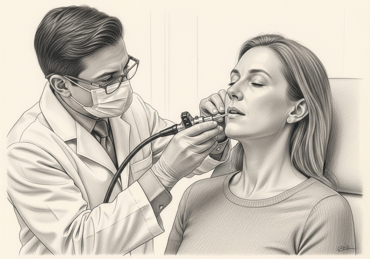
History and focused examination
The clinician documents the growth pattern, pain, prior skin cancers, radiation exposure, facial movement, tongue movement, sensation, oral findings, and neck nodes. The entire skin of the scalp, face, and ear may be examined when a parotid lesion is present. These details help distinguish a primary gland tumor from infection, a benign lesion, a lymph node, or metastasis from the skin. Clinical appearance alone cannot determine histology or grade.
Ultrasound
Ultrasound can define a superficial parotid or submandibular mass and guide needle sampling. It also evaluates nearby lymph nodes. It is efficient, avoids radiation, and can distinguish solid from cystic components. Deep-lobe, skull-base, or parapharyngeal extension may require CT or MRI.
Fine-needle aspiration or core biopsy
A needle sample is examined by a pathologist and may be categorized using a salivary cytology system. A core sample may provide additional architecture in selected cases. Preoperative tissue information helps plan the extent of surgery and counsel about nerve or neck management. Sampling can be indeterminate because salivary tumors are diverse and areas within one tumor may differ.
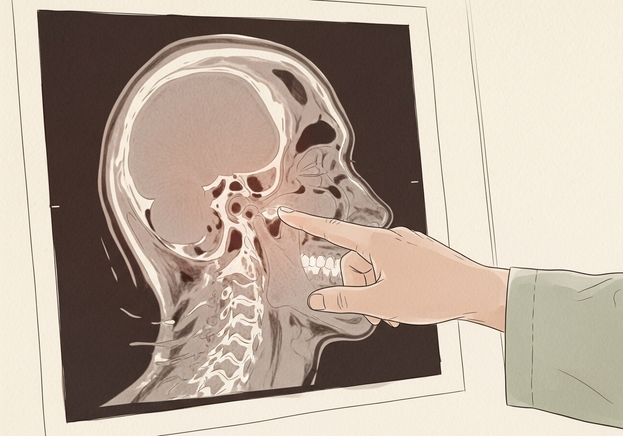
MRI or contrast-enhanced CT
MRI is useful for soft-tissue detail, deep-lobe disease, and suspected spread along nerves. CT can evaluate bone, neck anatomy, and the chest in appropriate cases. Imaging maps the tumor relative to the facial nerve region, skull base, vessels, jaw, throat, and lymph nodes. Imaging suggests behavior but does not replace pathology.
PET/CT and distant staging when indicated
Higher-grade, advanced, recurrent, or node-positive disease may warrant metabolic imaging or chest imaging to assess spread. The result can change whether surgery is curative, part of combined treatment, or not the preferred first step. Inflammation can produce false-positive uptake, and some slow-growing salivary cancers may show less activity.
Pathology and molecular review
The final diagnosis may require immunohistochemistry, fusion testing, receptor testing, or expert review. Accurate classification matters because grade, perineural invasion, margins, nodal disease, HER2, androgen receptor, or other features can influence treatment. Not every tumor needs broad molecular testing; it is selected according to histology and disease setting.
Understanding pathology, biomarkers, and staging
A complete pathology report identifies the histologic type, grade when applicable, tumor size and extent, surgical margins, lymphovascular invasion, perineural invasion, lymph-node findings, and extranodal extension. Because salivary tumors can look alike under the microscope, subspecialty review may be valuable when the diagnosis is rare or the biopsy and clinical behavior do not match.
Some salivary cancers are defined by characteristic molecular alterations. In recurrent or metastatic disease, testing for targets such as androgen receptor expression, HER2 amplification or overexpression, NTRK fusion, or other actionable findings may be considered according to tumor type and current guidelines. These results do not replace local treatment planning for a resectable tumor, but they can broaden options when disease is advanced.
Staging incorporates the gland or anatomic site, tumor size and extension, facial nerve or skin involvement, lymph nodes, and distant spread. The treating team should explain both the anatomic stage and the histology-specific features that influence risk.
Treatment planning
Treatment is individualized. A useful recommendation accounts for cancer control, expected speech and swallowing, airway safety, appearance, recovery time, medical fitness, prior treatment, personal priorities, and the possibility that more than one approach can be reasonable. The following options are discussed according to the diagnosis and stage.
Surgical removal of the primary tumor
Operations may include superficial, partial, or total parotidectomy; submandibular gland excision; resection of a sublingual or minor salivary tumor; or a larger composite operation when adjacent tissue is involved. The extent is chosen to obtain an appropriate margin while preserving uninvolved nerves and structures when possible.
Facial nerve management
When the facial nerve is not involved, preservation is a priority during parotid surgery. If a nerve segment is invaded by cancer, removal and immediate repair, grafting, nerve transfer, or other facial reanimation planning may be discussed. The decision depends on preoperative function, imaging, intraoperative findings, pathology, and the ability to obtain cancer control.
Neck dissection
Lymph-node surgery may be recommended for clinically involved nodes or selected high-risk tumors even when nodes are not obviously enlarged. Tumor grade, size, site, histology, and imaging help determine the appropriate neck levels.
Postoperative radiation therapy
Radiation may be recommended for high-grade disease, close or positive margins, perineural invasion, advanced stage, nodal disease, recurrent disease, or other higher-risk findings. The recommendation should be based on final pathology and reviewed with a radiation oncologist experienced in head and neck cancer.
Systemic and targeted therapy
For unresectable, recurrent, or metastatic disease, treatment may include clinical trials, targeted therapy for selected molecular findings, hormonal approaches for androgen-receptor–positive disease, HER2-directed therapy, chemotherapy, or immunotherapy in selected settings. Because salivary cancers are uncommon and biologically diverse, therapy should be matched to histology, biomarkers, prior treatment, disease pace, and patient goals.
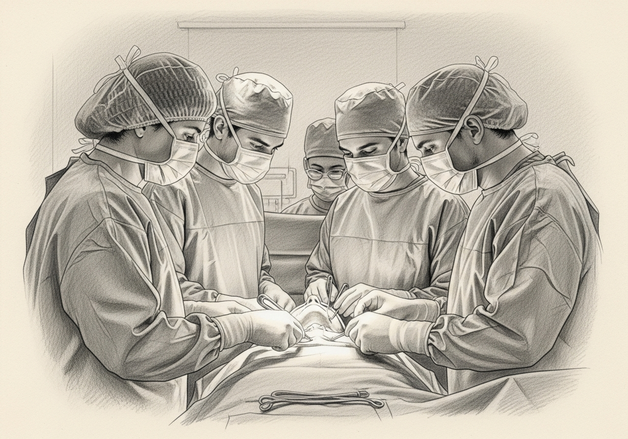
Surgical considerations
The operative plan begins with the gland and extends to every structure the tumor touches. In parotid surgery, the facial nerve is identified and followed through the gland. The surgeon may remove only the portion of gland containing the tumor or more extensive tissue when the cancer is large, deep, recurrent, or invasive. A neck dissection can be performed through the same or an extended incision when indicated.
If cancer directly invades a facial nerve branch, leaving involved nerve behind may compromise local control. The surgeon should discuss before the operation whether nerve sacrifice is possible, what reconstructive options are available, and how eye protection and facial movement would be addressed. Immediate cable grafting, masseteric or hypoglossal nerve transfer, static suspension, eyelid procedures, and staged reanimation are different tools selected according to the defect and prognosis.
Minor salivary cancers can require resection of palate, jaw, tongue, pharynx, or sinonasal structures. The planned margin must be balanced with speech, swallowing, dental rehabilitation, and reconstruction. For adenoid cystic carcinoma and other tumors prone to perineural spread, MRI and pathology are particularly important in defining the route toward the skull base.
Function, reconstruction, and rehabilitation
The functional impact depends on the gland and operation. Parotid surgery can affect facial movement, ear or cheek sensation, contour, and sweating over the cheek while eating. Submandibular surgery can affect lower-lip movement, tongue sensation, or tongue motion. Oral and palatal resections can alter speech, chewing, swallowing, and the separation between the mouth and nose.
Reconstruction may involve local tissue rearrangement, fat or muscle transfer, regional flaps, free tissue transfer, nerve grafting, facial reanimation, dental prosthetics, or an obturator. The best plan is defect-specific. A small surface defect may need little reconstruction, while a composite jaw or palate defect may require bone and soft tissue from another part of the body.
Speech-language pathology, nutrition, dental oncology, physical therapy, and facial rehabilitation can be incorporated before and after treatment. Functional planning should begin before the operation rather than after a problem becomes established.
Preparing for treatment
Preparation is not only a list of preoperative tests. It is an opportunity to identify problems that can make treatment harder and to establish a baseline for recovery. Depending on the plan, preparation may include:
- Pathology and imaging review: Bring the biopsy report and slides or blocks when requested, plus original ultrasound, CT, MRI, or PET images and reports.
- Baseline facial nerve documentation: Photographs, video, and a detailed facial movement examination establish whether weakness existed before surgery.
- Skin examination: Patients with a parotid-area node and prior skin cancer may need review of dermatology records and a complete scalp, face, and ear examination.
- Dental, swallowing, or nutrition assessment: These are considered when the tumor or planned treatment involves the mouth, palate, jaw, throat, or postoperative radiation.
- Medical optimization: Review anticoagulants, diabetes, cardiac and lung conditions, tobacco use, and prior radiation or surgery.
- Reconstructive planning: If nerve, skin, bone, or mucosal loss is possible, discuss reconstructive options and donor sites before the day of surgery.
- Support and logistics: Plan transportation, help at home, time away from work, drain care, eye care if facial weakness is possible, and attendance at pathology follow-up.
Patients should bring a complete medication list, allergy history, prior operative reports, pathology reports, imaging discs or secure links, and the names of the clinicians already involved. A written list of questions and a trusted support person can make a complex visit easier to absorb.
Recovery and what follow-up may involve
Recovery varies substantially because a small transoral procedure, a neck dissection, a major open resection, and combined surgery with reconstruction are very different experiences. The treating team should give procedure-specific instructions. A general framework is:
- Immediately after surgery: The team monitors facial movement or other relevant nerves, bleeding, airway, pain, swallowing, and drains. Larger operations may require a monitored unit.
- First one to two weeks: Swelling and bruising begin to improve. Drains and sutures are removed according to output and healing. Numbness and stiffness are common early effects.
- Pathology review: Final pathology determines whether margins are adequate and whether neck treatment, radiation, or systemic consultation is recommended.
- First several months: Facial nerve recovery, shoulder motion, speech, swallowing, and scar maturation are assessed. Rehabilitation is added when deficits are present.
- Long-term: Surveillance continues because some salivary cancers can recur late. Imaging and chest follow-up are individualized by histology and stage.
New or worsening breathing difficulty, brisk bleeding, rapidly increasing swelling, inability to manage saliva, signs of dehydration, chest pain, or a sudden neurologic change require urgent medical attention. Routine online messages are not appropriate for emergencies.
Risks and uncertainties to discuss
No treatment is risk-free, and risk changes with anatomy, extent of disease, prior radiation, nutrition, tobacco exposure, medical conditions, and the specific operation or nonsurgical regimen. Topics that may need discussion include:
- Bleeding, infection, fluid collection, or salivary leak: These can require observation, drainage, medication, pressure dressings, or another procedure.
- Temporary or permanent nerve weakness: Facial, lower-lip, tongue-movement, or tongue-sensation nerves can be stretched, injured, or intentionally removed if invaded by cancer.
- Eye exposure: Facial weakness can impair eyelid closure and requires lubrication, protection, and sometimes a procedure.
- Frey syndrome and contour change: Sweating or flushing over the cheek during meals and a hollowed contour can occur after parotidectomy.
- Swallowing, speech, or dental effects: These depend on oral, palatal, jaw, or throat involvement and the reconstruction used.
- Positive or close margins: Microscopic tumor can extend beyond what is visible, especially with perineural spread, leading to additional treatment recommendations.
- Recurrence or distant spread: Risk varies greatly by histology, grade, stage, and pathology, and may persist for years.
- Reconstructive complications: Graft, flap, or nerve repair problems can require additional procedures or prolonged rehabilitation.
This list is educational rather than a substitute for a consent discussion. The clinician should explain which risks are common, which are uncommon but serious, how they are reduced, and what alternatives exist in the patient's specific case.
Surveillance and survivorship
Follow-up serves several purposes: confirming healing, reviewing pathology, detecting recurrence, managing treatment effects, and helping patients return to daily life. A survivorship plan may address:
- Incision and nerve checks: Early visits assess healing, facial or tongue function, eye protection, drains, and infection.
- Pathology-directed planning: The surgical, radiation, and medical oncology teams review high-risk findings and adjuvant options.
- Imaging surveillance: MRI, CT, chest imaging, or other studies are selected by histology, stage, symptoms, and time from treatment.
- Rehabilitation: Facial therapy, speech and swallowing therapy, shoulder therapy, and scar or lymphedema management are added when needed.
- Dental and oral care: Patients with oral surgery or radiation need long-term prevention and monitoring.
- Skin cancer surveillance: Patients whose parotid or neck disease is related to cutaneous cancer require coordinated dermatologic follow-up.
- Late recurrence awareness: Some salivary histologies can recur many years later, so follow-up may extend beyond the typical early surveillance period.
The schedule is individualized by tumor site, stage, treatment, symptoms, and current guidelines. Patients should report new persistent symptoms between scheduled visits rather than waiting for the next appointment.
Getting a second opinion
A second opinion is particularly useful when the biopsy is indeterminate, the histology is rare, facial nerve sacrifice has been proposed, the tumor is recurrent, the disease tracks toward the skull base, or the treatment plan would combine major surgery and radiation. The review should include the actual pathology material and original imaging.
A meaningful second opinion does more than name an operation. It explains the certainty of the diagnosis, the expected margin, whether facial nerve preservation is oncologically reasonable, the need for neck dissection or radiation, reconstructive options, and what alternatives exist. Because salivary cancers are uncommon, a pathology or multidisciplinary review can materially change the plan in selected cases.
Questions to ask at a consultation
- What specific salivary cancer type and grade was found, and should the pathology be reviewed?
- Is the tumor arising in the gland itself or in a lymph node from skin cancer?
- Does imaging suggest facial nerve, skull-base, bone, skin, or deep-lobe involvement?
- What operation is recommended, and what part of the gland will be removed?
- What is the likelihood that a facial nerve branch will need to be removed?
- If nerve removal is necessary, what immediate and staged reconstruction options are available?
- Do I need a neck dissection?
- Which pathology findings would lead to postoperative radiation?
- Would molecular testing affect treatment now or if disease recurs?
- How will the plan affect facial movement, swallowing, speech, appearance, and return to work?

Request a consultation
If you have a salivary gland cancer diagnosis, an enlarging parotid or submandibular mass, facial weakness, or a treatment plan you want reviewed, request a Head & Neck consultation or call (212) 444-8006. Bring your pathology and original imaging when available. Seek emergency care for breathing difficulty, uncontrolled bleeding, or rapidly progressive swelling.
Specialists who treat salivary gland cancer
Dr. Moustafa Mourad
MD, FACS
Double Board-Certified Head & Neck and Facial Plastic & Reconstructive Surgeon
Dr. Moustafa Mourad is a double board-certified head and neck and facial plastic and reconstructive surgeon who cares for the full range of cosmetic and complex conditions affecting the face, head, and neck.
- Facial plastic and reconstructive surgery
- Head and neck cancer surgery
- Microvascular free-flap reconstruction
- Facial trauma and reconstruction
Also caring for this area

Not sure who to see? Our patient coordination team can help match you with the right specialist.
(212) 444-8006Frequently Asked Questions
No. Many salivary masses, especially in the parotid, are benign. Persistent or enlarging masses still require evaluation because examination alone cannot establish the diagnosis.
Fine-needle aspiration and appropriately selected core biopsy are standard diagnostic tools. The clinician chooses the technique and route to obtain useful tissue while minimizing risk.
New weakness is concerning and requires prompt assessment, but it is not proof of cancer. The pattern, imaging, and biopsy determine the cause.
When the nerve is functioning and not invaded, preservation is a priority. If cancer directly involves a branch or the main trunk, removal may be necessary to achieve an appropriate cancer operation. The decision is individualized.
Perineural invasion means tumor cells are seen around or within a nerve on pathology. It can influence staging, imaging, margin assessment, radiation recommendations, and surveillance.
Not every patient does. Radiation is more likely to be discussed for high-grade tumors, advanced stage, close or positive margins, nerve involvement, lymph-node disease, recurrence, or other high-risk findings.
Radiation or systemic therapy may be used when a tumor cannot be removed, when surgery would cause unacceptable harm, for recurrent or metastatic disease, or in selected individual situations. Many localized resectable cancers are treated primarily with surgery.
It removes both superficial and deep portions of the parotid gland. The term does not automatically mean removal of the facial nerve; nerve management depends on tumor involvement.
The schedule depends on histology and stage. Some salivary cancers can recur late, so follow-up may continue for many years.
It can be valuable for rare histology, indeterminate biopsy, discordant findings, or when treatment depends on grade or a specific molecular diagnosis.
Clinical References
These independent resources from medical and professional organizations offer further reading. They are provided for general education and do not replace a consultation with a clinician.
Related Resources
Related Procedures
Related Conditions

Request a consultation for salivary gland cancer
Schedule an evaluation with our team to review your symptoms and the appropriate next steps.