
About the Procedure
A cerebrospinal fluid (CSF) leak occurs when a defect in the skull base allows fluid from around the brain to drain into the nose or sinuses. Successful repair depends on confirming that the fluid is CSF, locating the defect, understanding why it occurred, and building a closure appropriate to its size, flow, and surrounding anatomy.
Most leaks can be repaired endoscopically through the nostrils, without external incisions or an open skull approach. Spontaneous leaks also require attention to pressure factors that can affect recurrence, and skull-base care is coordinated with neurosurgery when appropriate.
What this evaluation should clarify
A focused evaluation should answer a few key questions before any procedure is recommended:
- Is the underlying diagnosis and treatment goal established with the right examination, imaging, testing, or pathology?
- How does this option compare with continued medical care, a different procedure, observation, or referral to another specialty?
- Which anatomy, prior treatment, other health conditions, benefits, risks, and recovery requirements change the recommendation?
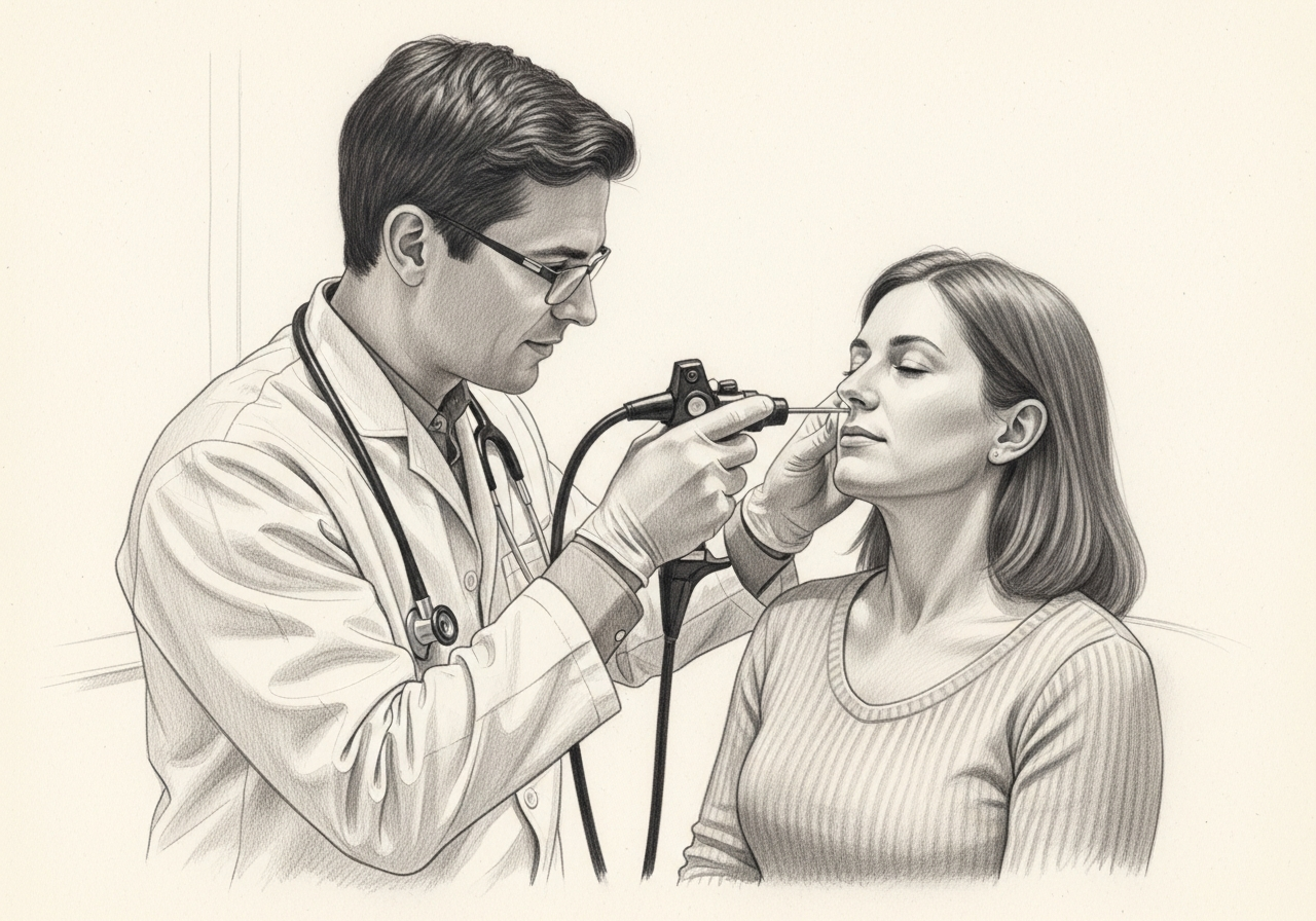
Considering endoscopic CSF leak repair? The next step is a quiet, unhurried conversation.
Evaluation and treatment pathway
The pathway is individualized and generally follows these steps:
- Confirm the diagnosis, the treatment goal, and the reasons a less invasive or nonsurgical approach is not enough.
- Gather laboratory and imaging evidence when it can be obtained, localize the defect, review any trauma or prior surgery and meningitis history, and assess for an associated encephalocele or pressure-related risk.
- Consider alternatives, which depend on the cause and may include monitored conservative care for selected acute traumatic leaks, a different surgical route, combined repair, or treatment of underlying intracranial-pressure factors.
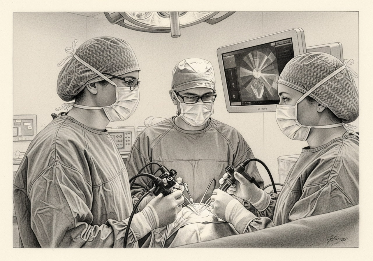
- During repair, the defect is exposed endoscopically, herniated tissue or surrounding bone is managed as appropriate, and the opening is reconstructed in layers; lumbar drainage, graft choice, and postoperative restrictions are individualized.
- Recovery includes clear instructions about activity, straining, nasal care, medications, follow-up, and the signs of a recurrent leak or meningitis, and long-term pressure evaluation may be part of care for spontaneous leaks.
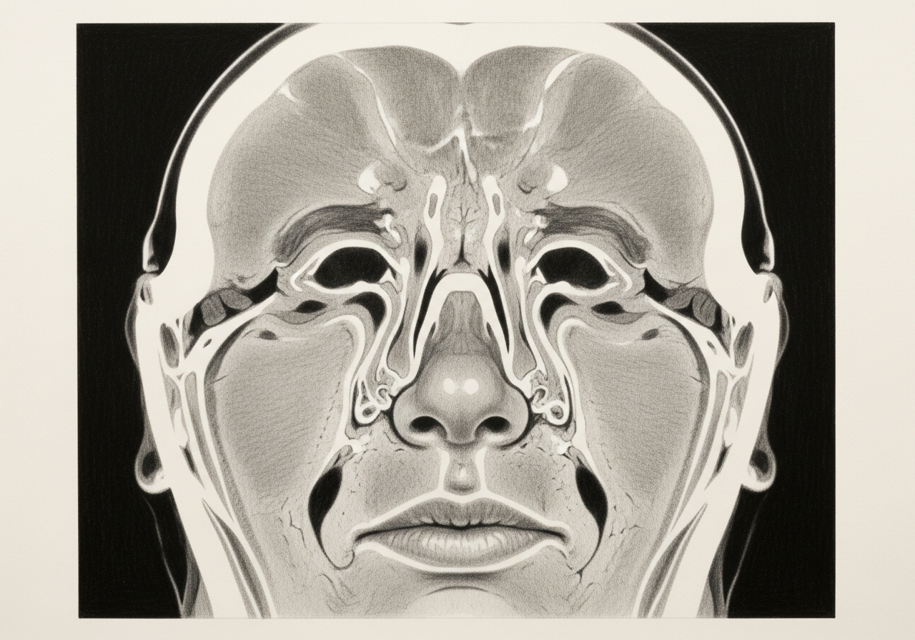
How it is performed
The surgeon uses endoscopes passed through the nostrils to locate the skull-base defect, often with image guidance. The area is prepared and the defect is sealed with grafts, which may include tissue from the nasal lining, along with supporting materials.
In most cases there are no external incisions and no need to open the skull. When elevated intracranial pressure is contributing, additional management may be planned with neurosurgery.
Risks and alternatives
Possible risks include recurrent or persistent leak, bleeding, infection including meningitis, and the need for additional surgery. When elevated intracranial pressure is involved, ongoing management is often needed to protect the repair.
Because an untreated CSF leak carries a meningitis risk, observation alone is usually not a safe long-term alternative, although the timing and approach are individualized.

Results and follow-up
Endoscopic repair is intended to seal the leak and restore the barrier between the nose and the brain. Some leaks, particularly those related to elevated intracranial pressure, may require further treatment to prevent recurrence.
Follow-up monitors for recurrent drainage and confirms healing, and ongoing care addresses any underlying contributing factors.
What to bring to your consultation
Gathering the right records ahead of time helps make the consultation productive. Useful items include:
- Imaging files and reports, such as prior CT or MRI scans
- Endoscopy or operative findings from earlier care
- Pathology results, if any
- Relevant laboratory results
- Notes from prior treatment
- A current medication list
- The specific question you want answered
When to seek urgent care
Some symptoms require urgent or emergency assessment, including fever, neck stiffness, a severe headache, confusion, clear recurrent drainage, vision changes, seizure, new weakness, major bleeding, or rapidly worsening illness. A routine appointment request or online form is not an emergency service; for emergency symptoms, seek immediate care.
Medical review
This page is a patient-education resource reviewed by the responsible Norelle ENT clinician before publication. It does not replace an in-person evaluation. If symptoms are severe or rapidly worsening, seek immediate medical care.
Specialists who perform endoscopic CSF leak repair
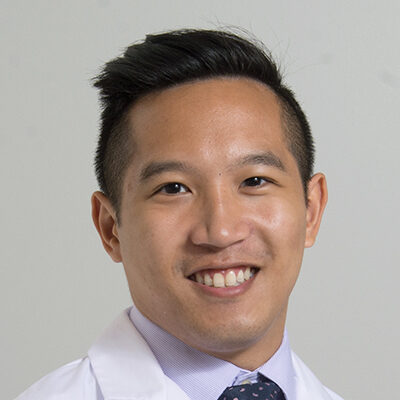
Dr. Adrian Ong
MD
Board-Certified Facial Plastic & Reconstructive and Head & Neck Surgeon
Dr. Adrian Ong is a board-certified surgeon who practices exclusively on the face, head, and neck, with expertise spanning rhinoplasty, sinus surgery, facial trauma, reconstruction, and sleep surgery.
- Functional and aesthetic rhinoplasty (including revision)
- Sinus surgery and complex revision sinus surgery
- Facial trauma and nasal fractures
- Head and neck cancer surgery and microvascular reconstruction
Also caring for this area


Not sure who to see? Our patient coordination team can help match you with the right specialist.
(212) 444-8006Frequently Asked Questions
Candidacy requires laboratory and imaging evidence when it can be obtained, localization of the defect, review of any trauma or prior surgery, meningitis history, and assessment for an associated encephalocele or pressure-related risk.
The defect is exposed endoscopically, herniated tissue or surrounding bone is managed as appropriate, and the opening is reconstructed in layers. Lumbar drainage, graft choice, and postoperative restrictions are individualized rather than routine for every case.
Alternatives depend on the cause and may include monitored conservative care for selected acute traumatic leaks, a different surgical route, combined repair, or treatment of underlying intracranial-pressure factors.
Patients need clear instructions about activity, straining, nasal care, medications, follow-up, and the signs of a recurrent leak or meningitis. Long-term pressure evaluation may be part of care for spontaneous leaks.
Clinical References
These independent resources from medical and professional organizations offer further reading. They are provided for general education and do not replace a consultation with a clinician.
Related Conditions

Related Procedures

1 of 2 · Skull Base Reconstruction
Request a consultation about endoscopic CSF leak repair
Schedule a consultation with our team to discuss whether this procedure is the right option for you.