
About the Procedure
Endoscopic endonasal skull-base surgery reaches selected lesions through the nose, avoiding some external incisions but not eliminating the complexity or risk of operating beside the brain, optic nerves, carotid arteries, pituitary, and cranial nerves. This page explains who is on the team, why this route may be favored for a given lesion, and what reconstruction and follow-up are required.
The skull base is the bony floor that separates the nose and sinuses from the brain and the structures around the eyes. Tumors and other lesions in this region were historically reached through open approaches, but many can now be accessed endoscopically through the nose and sinuses, typically by a team that includes an ENT skull-base surgeon and a neurosurgeon.
What this evaluation should clarify
Before recommending endoscopic skull base surgery, an evaluation should clarify a few core questions:
- Is the underlying diagnosis and treatment goal established with the right examination, imaging, testing, or pathology?
- How does this option compare with continued medical care, a different procedure, observation, or referral to another specialty?
- Which anatomy, prior treatment, comorbidities, benefits, risks, and recovery requirements change the recommendation?
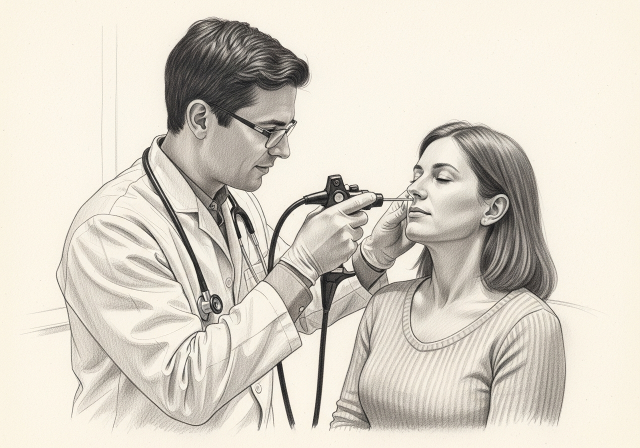
Considering endoscopic skull base surgery? The next step is a quiet, unhurried conversation.
Evaluation and treatment pathway
- Confirm the diagnosis, treatment goal, and the reasons a less invasive or nonsurgical approach is insufficient.
- Candidacy depends on the diagnosis, compartment, extension, vascular and neural relationships, prior treatment, and imaging quality, and on whether endonasal access provides an appropriate route compared with open, combined, radiation, or observation options.
- Alternatives include observation, medication for selected pituitary tumors, radiation, open cranial or facial approaches, combined approaches, and nonsurgical care. Team expertise and facility support are part of candidacy.
- ENT and neurosurgical roles vary by case. The approach creates a nasal corridor, treats the lesion, and reconstructs the skull-base defect using tissue selected for the size and leak risk; pathology, navigation, and vascular planning may be involved.
- Recovery varies widely with the lesion and extent. Patients may need nasal care, endocrine and neurologic monitoring, activity restrictions, serial imaging, and coordinated follow-up for vision, hormones, CSF leakage, and disease control.
Who may be a candidate
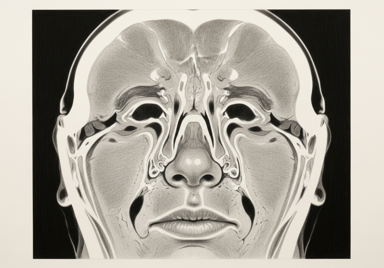
This approach may be considered for a range of skull-base conditions, including:
- Pituitary and other sellar tumors
- Certain sinonasal tumors extending to the skull base
- Some meningiomas and other lesions in reachable locations
- CSF leaks and skull-base defects
Not every lesion is suitable for an endoscopic approach. The decision depends on the location, the surrounding nerves and vessels, and the goals of treatment.
How it is performed
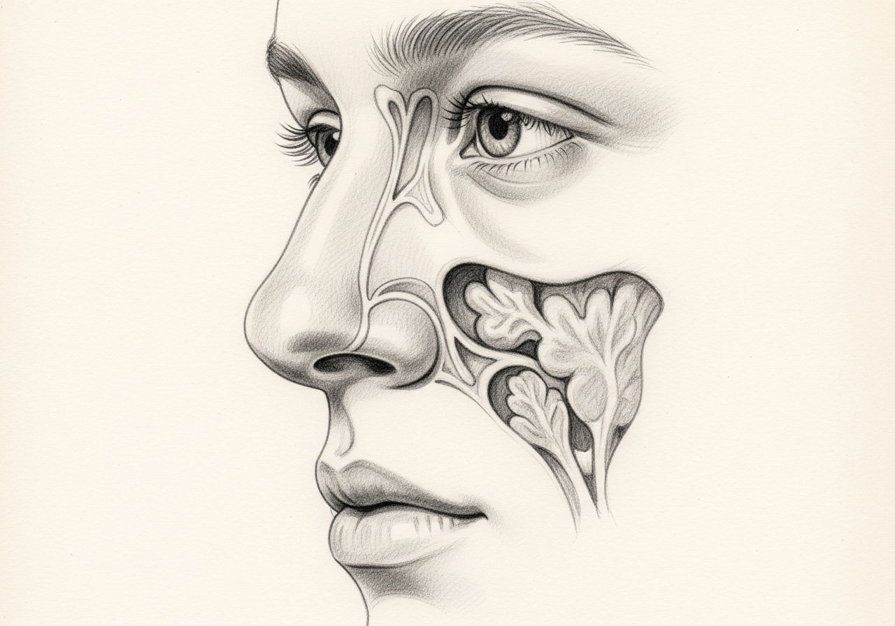
Endoscopes are passed through the nostrils, and the sinuses are opened to create a pathway to the skull base. Using image guidance, the team reaches the lesion and removes it while protecting the optic nerves, carotid arteries, and brain.
The skull base is then reconstructed, often using the patient's own tissue, to separate the nose from the space around the brain and reduce the risk of a CSF leak.
Risks and alternatives
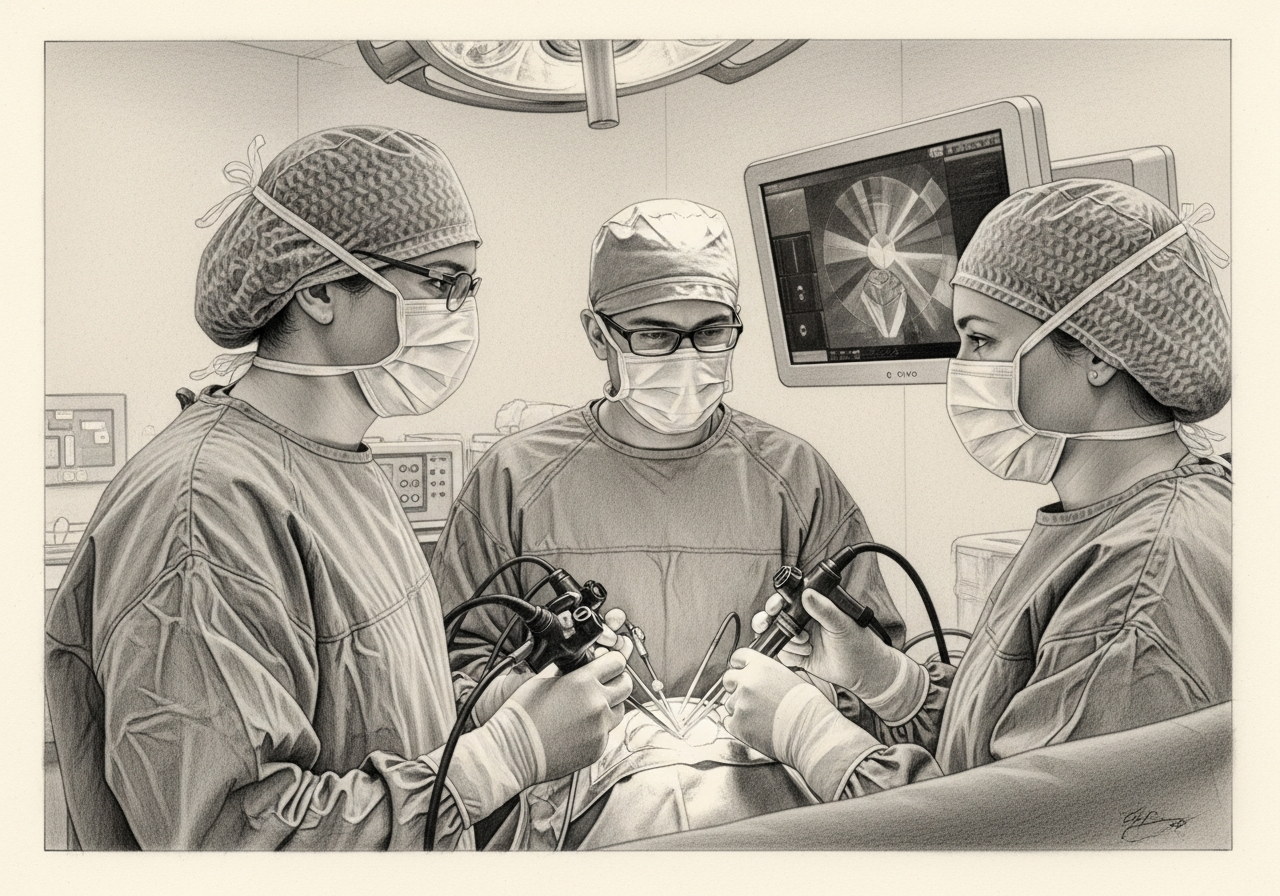
Possible risks include bleeding, infection, CSF leak, changes in smell, and, less commonly, injury to nerves or blood vessels near the skull base. The team takes specific steps to protect these structures and to reconstruct the skull base securely.
Alternatives depend on the diagnosis and may include observation, open surgical approaches, radiation therapy, or medical treatment. The approach is determined by the skull-base team.
Results and follow-up
The goals are to remove or control the lesion, protect nearby function such as vision, and restore the barrier between the nose and the brain. Some conditions require additional treatment after surgery.
Long-term follow-up with imaging and the relevant specialists monitors for recurrence and supports recovery.
What to bring to your consultation
Bring or securely transfer the records that can change this decision:
- Imaging files and reports
- Endoscopy or operative findings
- Pathology results
- Laboratory results
- Prior treatment notes
- A current medication list
- The specific question you want answered
Having these available helps the team review the diagnosis and the available options together.
When to seek urgent care
After surgery, seek urgent or emergency assessment for clear continuous drainage, fever with neck stiffness, severe headache, vision change, major bleeding, confusion, weakness, seizure, marked thirst or urination, faintness, or difficulty breathing.
An online form or routine appointment request is not an emergency service. For emergency symptoms, use emergency services rather than the routine form.
Medical review
This page is a patient-education resource reviewed by the responsible Norelle ENT clinician before publication. It does not replace an in-person evaluation. If symptoms are severe or rapidly worsening, seek immediate medical care.
Specialists who perform endoscopic skull base surgery
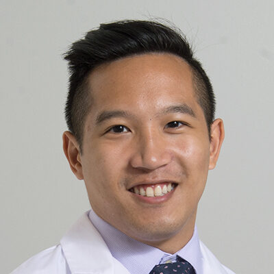
Dr. Adrian Ong
MD
Board-Certified Facial Plastic & Reconstructive and Head & Neck Surgeon
Dr. Adrian Ong is a board-certified surgeon who practices exclusively on the face, head, and neck, with expertise spanning rhinoplasty, sinus surgery, facial trauma, reconstruction, and sleep surgery.
- Functional and aesthetic rhinoplasty (including revision)
- Sinus surgery and complex revision sinus surgery
- Facial trauma and nasal fractures
- Head and neck cancer surgery and microvascular reconstruction
Also caring for this area

Not sure who to see? Our patient coordination team can help match you with the right specialist.
(212) 444-8006Frequently Asked Questions
Candidacy depends on diagnosis, compartment, extension, vascular and neural relationships, prior treatment, imaging quality, and whether endonasal access provides an appropriate route compared with open, combined, radiation, or observation options.
ENT and neurosurgical roles vary by case. The approach creates a nasal corridor, treats the lesion, and reconstructs the skull-base defect using tissue selected for the size and leak risk; pathology, navigation, and vascular planning may be involved.
Alternatives include observation, medication for selected pituitary tumors, radiation, open cranial or facial approaches, combined approaches, and nonsurgical care. Team expertise and facility support are part of candidacy.
Recovery varies widely with the lesion and extent. Patients may need nasal care, endocrine and neurologic monitoring, activity restrictions, serial imaging, and coordinated follow-up for vision, hormones, CSF leakage, and disease control.
Clinical References
These independent resources from medical and professional organizations offer further reading. They are provided for general education and do not replace a consultation with a clinician.
Related Conditions
Related Procedures

1 of 4 · Endoscopic Pituitary Surgery
Request a consultation about endoscopic skull base surgery
Schedule a consultation with our team to discuss whether this procedure is the right option for you.