
Overview
Congenital neck lesions may be noticed in childhood or appear later after an infection or growth. In an adult, however, a cystic neck mass should not be labeled congenital until malignancy has been appropriately excluded.
A congenital neck mass is a lump in the neck that arises from the way structures form before birth. Common types include thyroglossal duct cysts in the midline, branchial cleft cysts on the side of the neck, dermoid cysts, and lymphatic or vascular malformations. Many are noticed in infancy or childhood, but some are not recognized until adolescence or adulthood, sometimes after the mass becomes infected and suddenly swells.
Most congenital neck masses are benign. The reasons to evaluate one are to confirm what it is, to distinguish it from other causes of a neck lump, and to decide whether removal is advisable to prevent repeated infection or growth. In adults, a clinician will also confirm that a long-standing lump is genuinely congenital rather than a new problem.
Norelle ENT evaluates congenital neck masses with a careful history, examination, and imaging, and coordinates with radiology and pathology when needed. When surgery is appropriate, the plan is designed to remove the cyst and any associated tract completely while protecting nearby nerves, blood vessels, and glands.
How we approach the decision
Identifying the specific lesion, and excluding more serious causes in adults, guides everything that follows. The decisions this page is meant to help you understand are:
- Does the location and movement pattern fit a thyroglossal duct, branchial cleft, dermoid, vascular, or lymphatic lesion?
- Has imaging confirmed the anatomy and its relationship to the thyroid, hyoid bone, vessels, nerves, and airway?
- In an adult, has a metastatic cystic lymph node — especially HPV-related disease — been excluded?
Living with congenital neck mass? The next step is a quiet, unhurried conversation.
What happens next
Evaluation usually follows a clear sequence:
- An age-appropriate history and a location-based examination.
- Ultrasound and, when needed, cross-sectional imaging.
- Thyroid evaluation for midline lesions and targeted endoscopy for selected adult lateral cystic masses, since these can overlap with the workup for a neck mass evaluation.
- Fine needle aspiration or other tissue assessment when needed to exclude malignancy, including an unknown primary head and neck cancer or HPV throat cancer.
- Observation, infection control, sclerotherapy in selected vascular or lymphatic lesions, or definitive surgery. Midline thyroglossal lesions also require a thyroid nodule evaluation to confirm normal thyroid tissue before surgery.
Common types
The most frequent congenital neck masses include:
- Thyroglossal duct cyst: a midline cyst near the hyoid bone that often moves with swallowing or tongue movement
- Branchial cleft cyst: usually on the side of the neck, sometimes with a small skin opening
- Dermoid cyst: a firm cyst containing skin elements
- Lymphatic or vascular malformation: soft, sometimes compressible swellings present from early life
Each type behaves differently, which is why identifying the specific kind matters.
Symptoms and warning signs
Many congenital neck masses cause no symptoms and are noticed only as a lump. Others first appear when they become infected, causing pain, redness, and rapid swelling.
Features that deserve prompt attention include rapid enlargement, persistent drainage, signs of infection that do not settle, or any new firmness or fixation in an adult, since a neck lump in an adult should not be assumed to be congenital without evaluation.
Causes and risk factors
These masses result from normal embryologic structures that do not regress or fuse as expected. They are not caused by anything a parent or patient did.
There are usually no strong external risk factors. A family history is uncommon for most types. Infections of the upper respiratory tract can cause a previously silent cyst to swell and become noticeable.
How it is diagnosed
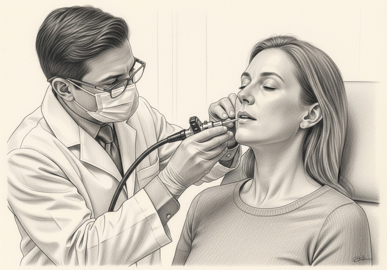
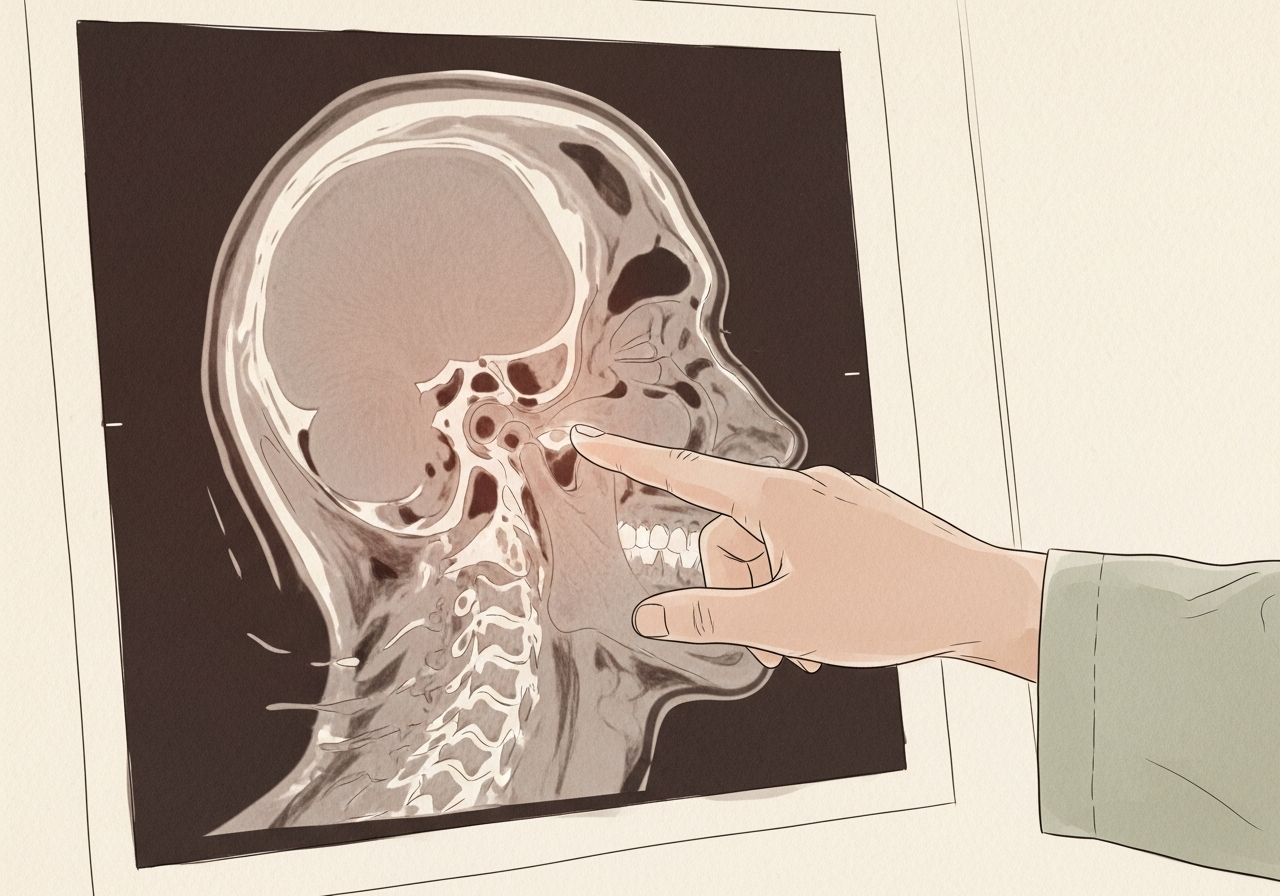
Evaluation begins with a history and a focused neck examination, including how the mass moves and where it sits. Imaging is the main diagnostic tool.
- Ultrasound is often the first study, especially in children
- CT or MRI may be used to map a tract or define deeper masses
- Fine needle aspiration can help when the diagnosis is unclear, particularly in adults
The aim is to confirm the type of mass and plan complete removal when surgery is indicated.
Treatment options
Treatment depends on the type of mass, symptoms, and whether infections have occurred. Small, stable, asymptomatic cysts can sometimes be observed.
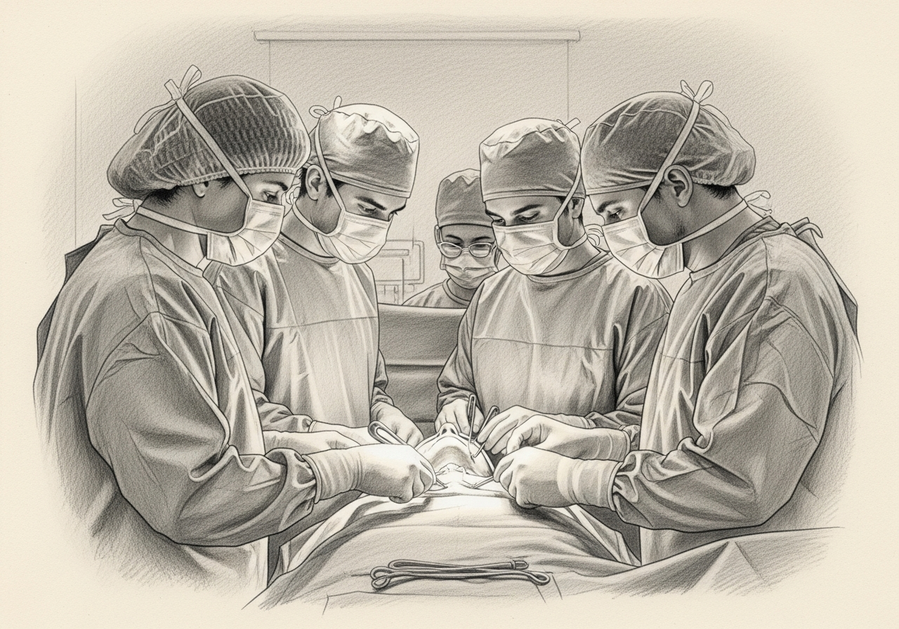
When a cyst causes repeated infections or continues to grow, surgical removal is often recommended. Active infection is usually treated first so the tissue is less inflamed at the time of surgery. Thyroglossal duct cysts are typically removed with a Sistrunk procedure, which removes the cyst along with the associated tract and a small portion of the hyoid bone to reduce the chance of recurrence.
When to seek urgent care
Use these categories to guide timing:
- Emergency, meaning call 911 or go to the nearest emergency department: rapid swelling with difficulty breathing or swallowing, or a severe spreading infection with high fever.
- Same-day or urgent evaluation: fever, spreading redness, or significant pain over the mass, or a quickly enlarging lump.
- Routine specialist evaluation: any persistent neck lump, recurrent swelling in the same area, or drainage from a small neck opening. In an adult, a cystic neck mass deserves timely specialist evaluation even when it looks benign on initial imaging.
The online consultation form is for routine scheduling and is not an emergency service.
Clinical perspective
Our head and neck surgeons distinguish midline from lateral lesions and apply a clear adult safety rule. A midline lump near the hyoid bone that moves with swallowing fits a thyroglossal duct cyst, while a lateral cystic lump fits a branchial cleft cyst in younger patients.
The most important factor is age. In an adult, a lateral cystic neck mass cannot be assumed to be a branchial cyst, because a cystic lymph-node metastasis from HPV-related oropharyngeal cancer can look similar and requires a different workup. For thyroglossal duct lesions, confirming normal thyroid tissue before surgery matters, and complete removal differs from a simple cyst excision.
What commonly changes the recommendation is the imaging anatomy and any tissue result. Candidacy for surgery, and the type of operation, is determined individually after specialist review rather than from the lump alone.
What to bring to your consultation
Bringing the right records makes a consultation more efficient. Helpful items include:
- Prior imaging and the written reports
- Pathology or biopsy results when available
- Recent laboratory results
- Treatment notes from any prior care
- A current medication list
- The specific decision you want the consultation to answer
Request a consultation for a focused review of the diagnosis, the available options, the likely tradeoffs, and the steps needed before treatment. For urgent symptoms, follow the guidance above rather than using the routine form.
Medical review
This page is a patient-education resource reviewed by the responsible Norelle ENT clinician before publication. It does not replace an in-person evaluation. If symptoms are severe or rapidly worsening, seek immediate medical care.
Specialists who treat congenital neck mass

Dr. Moustafa Mourad
MD, FACS
Double Board-Certified Head & Neck and Facial Plastic & Reconstructive Surgeon
Dr. Moustafa Mourad is a double board-certified head and neck and facial plastic and reconstructive surgeon who cares for the full range of cosmetic and complex conditions affecting the face, head, and neck.
- Facial plastic and reconstructive surgery
- Head and neck cancer surgery
- Microvascular free-flap reconstruction
- Facial trauma and reconstruction
Also caring for this area

Not sure who to see? Our patient coordination team can help match you with the right specialist.
(212) 444-8006Frequently Asked Questions
Most are benign cysts or developmental remnants. The main concerns are recurrent infection and growth, and in adults the need to confirm that a lump is truly congenital rather than a new problem. Evaluation clarifies the diagnosis.
Cystic lymph-node metastases, particularly from HPV-related oropharyngeal cancer, can look similar to a branchial cleft cyst and require a different workup. In an adult, malignancy is excluded before a lateral cystic mass is labeled congenital.
It is a midline developmental remnant near the hyoid bone that often moves with swallowing or tongue protrusion and can become infected. Removal is planned to take the cyst along with its tract and a central portion of the hyoid bone.
A cyst that has been quiet for years can swell after an upper respiratory infection, which makes it noticeable for the first time. Imaging and examination help confirm that the mass is a long-standing congenital cyst.
Not always. Small, stable, symptom-free cysts can sometimes be monitored. Removal is often recommended when a cyst causes repeated infections, grows, or remains uncertain after imaging.
It is the standard operation for a thyroglossal duct cyst. It removes the cyst along with its tract and a small central portion of the hyoid bone, which lowers the chance the cyst will come back.
Usually yes. Operating on actively inflamed tissue is more difficult, so an infection is generally treated first and surgery is planned once the area has settled.
A history, a neck examination, and imaging such as ultrasound, CT, or MRI are the main tools. Fine needle aspiration is sometimes added, especially in adults, to clarify the diagnosis.
Recurrence is uncommon when the cyst and any associated tract are removed completely. Incomplete removal or prior infection can increase the chance of recurrence, which is why the operation is planned carefully.
Clinical References
These independent resources from medical and professional organizations offer further reading. They are provided for general education and do not replace a consultation with a clinician.
Related Procedures

Related Conditions

Request a consultation for congenital neck mass
Schedule an evaluation with our team to review your symptoms and the appropriate next steps.