

Overview
Unknown primary head and neck cancer is diagnosed when malignant cells are found in one or more lymph nodes in the neck but the site where the cancer began is not identified during the initial evaluation. In most contemporary cases the pathology is squamous cell carcinoma. Some are associated with human papillomavirus and arise from a very small tumor hidden in the tonsil or base of tongue; others may be related to the skin, nasopharynx, or another mucosal site. The phrase “unknown primary” describes the diagnostic situation, not a single biological disease.
For patients, the experience can be unsettling because there is a confirmed cancer diagnosis without an obvious source. A careful workup is important because locating the primary tumor can narrow the area that needs treatment, clarify staging, and influence whether surgery, radiation, systemic therapy, or a combination is most appropriate. The evaluation should be organized, timely, and coordinated with pathology and radiology review.
How we approach the decision
This page is meant to help you understand the choices that organize an unknown-primary workup, then connect each one to the next appropriate step. Three questions usually frame the discussion:
- What does the node pathology suggest about the likely primary site—p16/HPV, EBV, thyroid, salivary, skin, or another origin? A persistent neck mass is often the starting point.
- Has the imaging and endoscopic examination been sequenced to maximize the chance of finding the primary, including a possible HPV-related oropharyngeal cancer hidden in the tonsil or base of tongue?
- Would targeted transoral robotic surgery or neck dissection change treatment fields or intensity? When the primary remains unclear, a head and neck second opinion can help confirm the plan.

Living with unknown primary head and neck cancer? The next step is a quiet, unhurried conversation.
Why this condition deserves a focused evaluation
A neck node can be the first and only visible sign of a small head and neck cancer. Treating only the enlarged node without understanding the likely source can leave untreated disease or expose a broader area than necessary to surgery or radiation. Conversely, repeating tests without a clear plan can delay care. A focused head and neck evaluation aims to confirm the pathology, determine whether the tumor is HPV- or Epstein–Barr virus related when relevant, map the involved lymph-node levels, and search the mucosal and skin surfaces that commonly drain to those nodes.
The workup also matters because “unknown” can change with better imaging, expert pathology review, examination under anesthesia, directed tonsil surgery, or transoral evaluation of the base of tongue. Even when no primary site is found, the pattern of nodal disease and biomarker results often guide a rational treatment field.
Anatomy and where the disease begins
Lymph nodes in the neck are arranged in defined levels that receive drainage from different parts of the scalp, face, mouth, throat, voice box, thyroid, salivary glands, and upper chest. A node high in the side of the neck has a different set of likely sources than a node near the collarbone. This drainage pattern is one of the first clues used by the treating team.
HPV-related squamous cell carcinoma commonly arises within lymphoid tissue of the palatine tonsil or the lingual tonsils at the base of tongue. These tumors can be very small and difficult to see while producing a relatively large cystic neck node. Epstein–Barr virus–associated disease may suggest the nasopharynx. A history of cutaneous squamous cell carcinoma can point toward skin of the scalp, face, or ear. The anatomy and pathology must therefore be interpreted together.
Symptoms and warning signs
Many patients first notice a painless lump in the side of the neck. The mass may fluctuate, feel cystic, or be mistaken for an infection. Other symptoms depend on the hidden site and may be absent.
- Persistent neck mass: A lump lasting more than two to three weeks, enlarging, or recurring after antibiotics deserves evaluation in an adult.
- One-sided throat or ear pain: Pain referred to one ear with a normal ear examination can arise from the tonsil, base of tongue, or another throat site.
- Swallowing change: A sensation of food sticking, pain with swallowing, or difficulty moving food through the throat may be relevant.
- Tonsil asymmetry or throat fullness: A subtle difference between the tonsils or a feeling of fullness may prompt targeted examination.
- Voice change, nasal symptoms, or skin lesion: These symptoms can point toward the larynx, nasopharynx, or a cutaneous source, although many patients have none of them.
- Unexplained weight loss or fatigue: These are nonspecific symptoms but should be considered in the broader clinical context.
Symptoms alone cannot establish the diagnosis. Benign infections, inflammation, reflux, dental disease, nerve problems, and other conditions can cause overlapping complaints. The purpose of evaluation is to understand the pattern, examine the relevant anatomy, and decide whether tissue sampling or imaging is appropriate rather than assuming the most serious or the most reassuring explanation.

When to seek urgent care
Most neck masses do not require an emergency department visit, but certain symptoms indicate that waiting for a routine appointment may be unsafe.
- Difficulty breathing, noisy breathing at rest, or a rapidly narrowing airway
- Brisk bleeding from the mouth or throat, coughing up a significant amount of blood, or bleeding that does not stop
- Rapidly expanding neck swelling, especially after a biopsy or procedure
- Inability to swallow liquids or manage saliva, signs of dehydration, or sudden severe weakness
Causes and risk factors
Unknown-primary disease is not caused by a single risk factor. The workup considers exposures and prior diagnoses that shift the probability toward particular primary sites.
- High-risk HPV: HPV-related cancers frequently involve the tonsil and base of tongue and can occur in people without a tobacco history.
- Tobacco and alcohol: These exposures increase the risk of several mucosal head and neck squamous cell cancers.
- Prior skin cancer: Cutaneous squamous cell carcinoma of the scalp, face, or ear can spread to parotid or neck nodes.
- Epstein–Barr virus and ancestry: These can be relevant when nasopharyngeal cancer is suspected.
- Prior radiation or prior cancer treatment: Treatment history can change both the likely diagnosis and the feasibility of future surgery or radiation.
- Immune suppression: Transplant medications and some immune disorders increase the risk and aggressiveness of cutaneous squamous cell carcinoma.
A risk factor changes probability; it does not prove that a person has cancer, and the absence of a recognized risk factor does not rule it out. A clinician uses risk information to determine the urgency and breadth of the workup, not as a substitute for examination and diagnosis.
How the evaluation is performed
A complete workup is assembled in steps. Not every person needs every test, and the order can change when there is airway compromise, significant bleeding, a rapidly enlarging mass, or a prior pathology diagnosis. The aim is to answer three separate questions: what is the abnormality, where did it begin, and how far does it extend?
History and complete head and neck examination
The clinician reviews the timeline, prior infections, tobacco and alcohol exposure, HPV-related history, skin cancers, previous biopsies, and prior treatment. The examination includes the scalp, skin, oral cavity, oropharynx, salivary glands, thyroid, and the pattern of neck nodes. This establishes which primary sites are most plausible and whether there are signs that require urgent airway or bleeding management. A normal office examination does not exclude a small tumor hidden in tonsil tissue or deep mucosa.
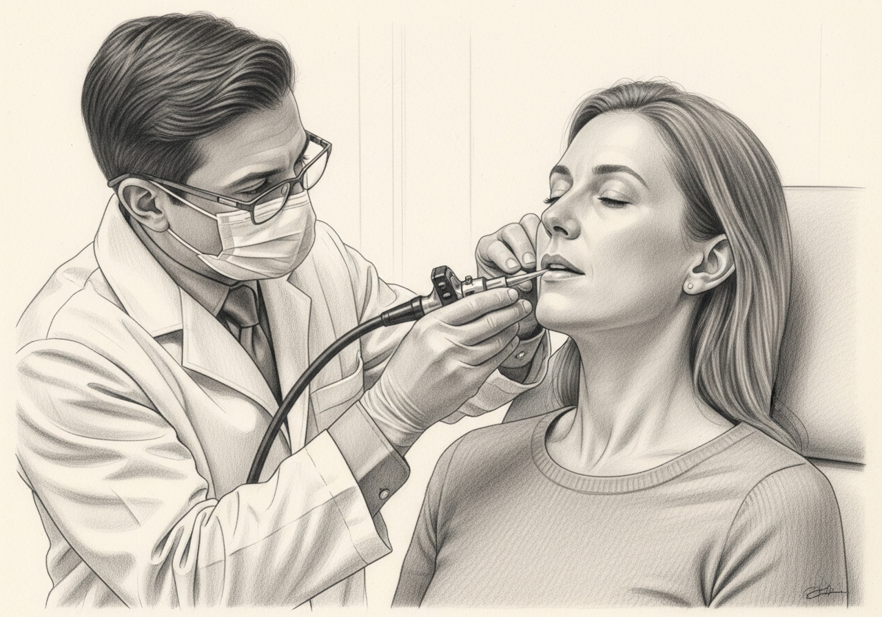
Flexible nasopharyngolaryngoscopy
A slender camera is passed through the nose after topical preparation to inspect the nasopharynx, base of tongue, throat, and larynx. It allows direct assessment of areas not visible through the mouth and can identify asymmetry or mucosal changes that guide biopsy. Very small or submucosal tumors can still be missed, so additional evaluation may be needed.
Ultrasound and needle biopsy
Ultrasound can characterize the node and guide fine-needle aspiration or core biopsy. The sample is reviewed to identify the tumor type and, when appropriate, HPV-related markers, EBV testing, or other studies. A tissue diagnosis is essential before definitive treatment, and biomarker results can focus the search for the primary site. Open excision of a suspicious node is generally avoided as the first diagnostic step when a needle biopsy can establish the diagnosis.
Cross-sectional imaging
Contrast-enhanced CT or MRI maps the involved nodes and looks for a primary tumor. MRI can be particularly helpful for soft-tissue and skull-base detail, while CT is efficient for the neck and chest. Imaging helps stage the disease and plan surgery or radiation. Inflammation, dental artifact, and very small tumors can limit interpretation.
PET/CT when indicated
Metabolic imaging can identify a subtle primary site, additional nodes, distant disease, or a separate cancer. It is often most useful before biopsies or tonsil surgery create inflammatory changes that may complicate interpretation. PET uptake is not specific for cancer and must be correlated with examination and pathology.
Examination under anesthesia and directed surgery
The surgeon may inspect and palpate the throat under anesthesia, biopsy suspicious areas, remove the palatine tonsils, and evaluate or remove lingual tonsil tissue using transoral techniques in selected cases. These steps can reveal a small primary site and may reduce the amount of tissue that needs radiation. The extent should be individualized; broader procedures add bleeding, pain, airway, and swallowing considerations.
Understanding pathology, biomarkers, and staging
The pathology report should identify the tumor type and may include p16 immunohistochemistry or direct HPV testing when an oropharyngeal origin is suspected. HPV-related nodal disease is staged differently from HPV-negative disease because the biology and prognosis differ. EBV testing may support a nasopharyngeal source. Keratinization, extranodal extension, number and size of nodes, laterality, and prior skin-cancer history also matter.
A second pathology review can be valuable when the sample is small, the diagnosis is unusual, or treatment will differ based on the result. Staging is not simply the size of the visible lump. It integrates the nodal levels, number of nodes, laterality, extranodal extension, biomarker status, imaging, and whether a primary site is eventually identified. The final stage and treatment plan should be explained in plain language.
Treatment planning
Treatment is individualized. A useful recommendation accounts for cancer control, expected speech and swallowing, airway safety, appearance, recovery time, medical fitness, prior treatment, personal priorities, and the possibility that more than one approach can be reasonable. The following options are discussed according to the diagnosis and stage.
Surgery to identify and treat the likely source
Selected patients undergo transoral evaluation or removal of tonsil and base-of-tongue tissue, often combined with neck dissection. Finding and removing a small primary may provide definitive pathology and can help tailor postoperative treatment. The decision depends on node burden, tumor accessibility, expected function, prior treatment, and whether surgery is likely to avoid or reduce additional therapy.
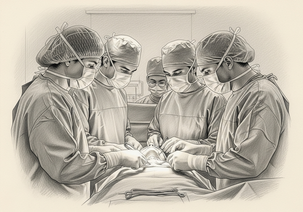
Neck dissection
Surgery can remove lymph nodes from defined neck levels, clarify the extent of disease, and treat gross nodal cancer. The type of neck dissection is based on the location and burden of disease, and the operation may be combined with transoral surgery.
Radiation therapy
Radiation can treat the involved neck and the mucosal sites considered at risk. Modern planning attempts to limit dose to uninvolved structures while maintaining cancer control. The field and dose depend on whether a primary is identified, HPV or EBV status, nodal stage, pathology, and prior treatment.
Systemic therapy with radiation
Chemotherapy or another systemic agent may be recommended with radiation for selected higher-risk disease. The choice reflects stage, pathology, medical fitness, kidney and hearing function, and multidisciplinary recommendations.
Clinical trials and individualized approaches
Some patients may be eligible for trials evaluating treatment intensity, imaging, surgery, radiation volumes, or systemic therapy. A trial should be discussed in relation to standard options, potential benefits, burdens, and eligibility criteria.
Surgical considerations
Surgery can serve two different roles: locating the hidden primary and treating known disease in the neck. These goals should be separated during consent. A transoral tonsil or base-of-tongue procedure can create throat pain and bleeding risk even when the primary is not ultimately found. Neck dissection can cause numbness, tightness, shoulder weakness, lymphatic swelling, and a visible scar, although preservation of nerves, vessels, and muscles is prioritized when oncologically appropriate.
If the likely primary lies in the tonsil or base of tongue, the surgeon may discuss conventional transoral techniques, laser surgery, or robotic assistance depending on anatomy, equipment, and experience. Surgery is not automatically superior to radiation, and it does not guarantee that radiation will be unnecessary. The value of surgery lies in what it can accomplish for a particular patient: removal of disease, more precise staging, identification of the primary site, or reduction of uncertainty.
Function, reconstruction, and rehabilitation
Treatment can affect swallowing, saliva, taste, neck and shoulder motion, speech, and appearance. Baseline swallowing assessment is especially useful when the plan may include transoral surgery, bilateral neck treatment, or radiation. A speech-language pathologist can document current function, teach exercises, and evaluate coughing, choking, or weight loss. Nutrition support may be needed before, during, or after treatment.
Reconstruction is not required for every unknown-primary case, but it becomes relevant when resection is extensive, prior radiation has impaired tissue quality, or a skin or mucosal defect needs coverage. The treatment plan should address not only removal of cancer but also how the patient will eat, speak, work, and recover.
Preparing for treatment
Preparation is not only a list of preoperative tests. It is an opportunity to identify problems that can make treatment harder and to establish a baseline for recovery. Depending on the plan, preparation may include:
- Records collection: Obtain the pathology report, pathology slides or blocks when requested, all imaging with reports, prior operative notes, and a timeline of antibiotics or procedures.
- Dental and swallowing assessment: These may be appropriate before radiation or extensive surgery to reduce avoidable complications and establish a baseline.
- Medical optimization: Control blood pressure and diabetes, review anticoagulants, assess heart and lung risk, and address tobacco and alcohol use.
- Nutrition: Document recent weight change and consider dietitian input when swallowing is difficult or treatment is expected to interfere with eating.
- Treatment mapping: Ask the team to explain which sites and neck levels will be treated, what information is still uncertain, and how each possible pathology result would change the plan.
- Support planning: Arrange transportation, help at home, communication with work, and a person who can participate in complex appointments.
Patients should bring a complete medication list, allergy history, prior operative reports, pathology reports, imaging discs or secure links, and the names of the clinicians already involved. A written list of questions and a trusted support person can make a complex visit easier to absorb.
Recovery and what follow-up may involve
Recovery varies substantially because a small transoral procedure, a neck dissection, a major open resection, and combined surgery with reconstruction are very different experiences. The treating team should give procedure-specific instructions. A general framework is:
- First days: Pain control, hydration, airway observation, drain care, and early mobility are priorities after surgery. Throat procedures may cause significant pain with swallowing, and bleeding precautions are essential.
- First several weeks: The team reviews final pathology, removes drains or sutures as appropriate, monitors shoulder and neck motion, and decides whether radiation or systemic therapy is recommended.
- During adjuvant therapy: Nutrition, dental care, skin care, swallowing exercises, and symptom control become central. Patients should report dehydration, fever, uncontrolled pain, or inability to maintain intake.
- Longer-term recovery: Surveillance, rehabilitation, thyroid monitoring after neck radiation, management of dry mouth or lymphedema, and psychosocial support may continue for months or years.
New or worsening breathing difficulty, brisk bleeding, rapidly increasing swelling, inability to manage saliva, signs of dehydration, chest pain, or a sudden neurologic change require urgent medical attention. Routine online messages are not appropriate for emergencies.

Risks and uncertainties to discuss
No treatment is risk-free, and risk changes with anatomy, extent of disease, prior radiation, nutrition, tobacco exposure, medical conditions, and the specific operation or nonsurgical regimen. Topics that may need discussion include:
- Diagnostic uncertainty: Even a thorough evaluation may not identify the primary site. The plan must still treat the known disease and the most likely areas of origin.
- Bleeding and airway problems: Transoral biopsies or tonsil procedures can bleed, and swelling can affect the airway in selected cases.
- Swallowing and nutrition changes: Pain, edema, nerve effects, scar, and radiation can impair swallowing temporarily or persistently.
- Neck and shoulder effects: Neck dissection may cause numbness, stiffness, shoulder weakness, or lymphedema.
- Treatment overlap: Surgery can be followed by radiation or chemoradiation if pathology shows higher-risk features, resulting in combined-treatment side effects.
- Recurrence or a later-appearing primary: Ongoing surveillance is needed because disease can recur and an occult primary can occasionally become evident later.
This list is educational rather than a substitute for a consent discussion. The clinician should explain which risks are common, which are uncommon but serious, how they are reduced, and what alternatives exist in the patient's specific case.
Surveillance and survivorship
Follow-up serves several purposes: confirming healing, reviewing pathology, detecting recurrence, managing treatment effects, and helping patients return to daily life. A survivorship plan may address:
- Examination: Regular head and neck examination and endoscopy are used to assess the treated sites and investigate new symptoms.
- Imaging: Post-treatment imaging is individualized according to stage, treatment, and findings.
- Swallowing and nutrition: Weight, aspiration symptoms, diet, and rehabilitation needs should be reviewed.
- Dental and oral health: Patients receiving radiation need a coordinated prevention and follow-up plan.
- Thyroid function: Neck radiation can affect thyroid function, so blood testing may be recommended.
- Tobacco and alcohol support: Stopping tobacco and moderating or avoiding alcohol can reduce future health risks.
- Psychosocial health: Anxiety about recurrence, body image, work, relationships, and financial strain deserve direct attention.
The schedule is individualized by tumor site, stage, treatment, symptoms, and current guidelines. Patients should report new persistent symptoms between scheduled visits rather than waiting for the next appointment.
Getting a second opinion
A second opinion is reasonable when the primary site remains unclear, pathology or HPV results are uncertain, more than one treatment pathway is possible, or a major operation or broad radiation field has been proposed. The reviewing team should receive the actual imaging, pathology material, operative notes, and prior treatment records—not only a summary. A useful second opinion explains what is known, what remains uncertain, which additional tests could change management, and how surgery and nonsurgical treatment compare for cancer control and long-term function. Seeking another opinion should not create an open-ended delay; the clinicians should help define a safe timeline.
Questions to ask at a consultation
- What type of cancer was found in the lymph node, and has the pathology been reviewed?
- Were p16, HPV, EBV, or skin-cancer markers tested, and what do the results imply?
- Which primary sites are most likely based on the lymph-node level and imaging?
- Do I need PET/CT, examination under anesthesia, tonsillectomy, or lingual tonsil evaluation?
- Could surgery identify the primary and reduce the radiation field?
- Will I need a neck dissection, and which neck levels would be removed?
- What are the alternatives to surgery?
- What is the likelihood that postoperative radiation or chemoradiation will still be recommended?
- How might each option affect swallowing, shoulder function, saliva, and return to work?
- What records should I send for a second opinion, and how quickly should treatment begin?

Request a consultation
If cancer has been found in a neck lymph node and the original site is uncertain, a focused consultation can review the pathology, imaging, endoscopic findings, and proposed treatment plan. Request an appointment with the Head & Neck team or call (212) 444-8006. For breathing difficulty, uncontrolled bleeding, or rapidly worsening swelling, seek emergency care instead of using the online form.
Specialists who treat unknown primary head and neck cancer
Dr. Moustafa Mourad
MD, FACS
Double Board-Certified Head & Neck and Facial Plastic & Reconstructive Surgeon
Dr. Moustafa Mourad is a double board-certified head and neck and facial plastic and reconstructive surgeon who cares for the full range of cosmetic and complex conditions affecting the face, head, and neck.
- Facial plastic and reconstructive surgery
- Head and neck cancer surgery
- Microvascular free-flap reconstruction
- Facial trauma and reconstruction
Also caring for this area


Not sure who to see? Our patient coordination team can help match you with the right specialist.
(212) 444-8006Frequently Asked Questions
A very small tumor can shed cells into a lymph node before it becomes visible on examination or imaging. Tumors hidden within tonsil tissue can be particularly difficult to detect. Pathology and the pattern of lymph-node involvement often provide clues even when the source is not found.
No. The term refers to the missing original site, not necessarily to distant spread. Some patients have disease limited to one or more neck nodes. Imaging and staging determine whether disease is present elsewhere.
HPV-related squamous cell carcinoma commonly arises from the tonsil or base of tongue, has a distinct staging system, and can influence the diagnostic search and treatment discussion. HPV status is interpreted with the full pathology and clinical picture.
Removing an involved node alone may not address other microscopic disease or the hidden primary site. Definitive treatment is planned for the full pattern of risk and may include a formal neck dissection, transoral surgery, radiation, systemic therapy, or a combination.
A small primary can be hidden within tonsil tissue and may not be visible from the surface. The extent of tonsil surgery depends on the side of the neck disease, imaging, HPV status, and the treating team's approach.
Lingual tonsils are lymphoid tissue at the base of tongue. In selected patients, transoral removal of part of this tissue can identify an otherwise occult primary. The procedure has pain, bleeding, airway, and swallowing considerations and is not required for every patient.
Sometimes a more precise diagnosis and favorable surgical pathology can reduce treatment, but surgery does not guarantee that radiation will be unnecessary. Node burden, margins, extranodal extension, and other features determine postoperative recommendations.
A confirmed metastatic neck node deserves timely evaluation. The exact timeline depends on symptoms, airway safety, pathology, and scheduling of imaging or procedures. The team should avoid both unnecessary delay and poorly planned duplicate testing.
Review can be useful when the diagnosis is uncommon, the sample is limited, HPV or EBV testing is unclear, or treatment would change based on the interpretation. The treating team can advise whether an expert review is appropriate.
Bring the biopsy report and, if requested, slides or tissue blocks; original imaging and reports; prior operative notes; medication and medical history; and a concise timeline of symptoms and tests. These materials allow the reviewer to give a meaningful opinion rather than repeat the entire workup.
Clinical References
These independent resources from medical and professional organizations offer further reading. They are provided for general education and do not replace a consultation with a clinician.
Related Resources
Related Procedures

Related Conditions

Request a consultation for unknown primary head and neck cancer
Schedule an evaluation with our team to review your symptoms and the appropriate next steps.