
About the Procedure
Submandibular gland excision is surgery to remove the salivary gland that sits beneath the jaw on one side of the neck. It is most often performed for a tumor, for stones that repeatedly block the gland and cause swelling and infection, or for chronic inflammation that does not improve with other treatment.
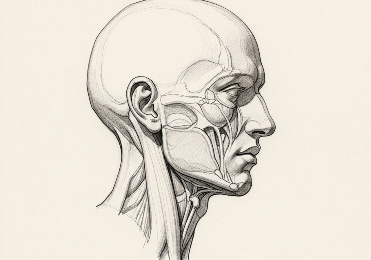
Several important nerves run near this gland, including the branch of the facial nerve that moves the lower lip, the nerve that provides sensation and taste to part of the tongue, and the nerve that moves the tongue. A central part of the operation is identifying and protecting these nerves.
Norelle ENT approaches submandibular gland excision with careful evaluation, often including imaging and a needle biopsy for a mass, and with attention to nerve preservation and a well-placed incision. The plan depends on the diagnosis, imaging, symptoms, and the patient's goals.
How we approach the decision
Submandibular gland surgery is planned around the cause of the problem and the nerves that surround the gland. A consultation helps work through several questions.
- Can the stone or duct problem be treated without removing the gland?
- Is a tumor diagnosis secure, and does cancer suspicion call for neck treatment or a wider operation?
- How will the marginal mandibular, lingual, and hypoglossal nerves be protected?
These questions are shaped by the broader picture of salivary gland disease and by how an adult neck mass is evaluated, along with what imaging and biopsy show.
Considering submandibular gland excision? The next step is a quiet, unhurried conversation.
What happens next
Care usually follows a clear sequence.
- Define the cause and anatomy with examination and imaging.
- Consider transoral stone removal, sialendoscopy, or other gland-preserving options through salivary gland surgery when appropriate and available.
- Plan the neck incision, gland and duct control, nerve identification, and tumor margins.
- Manage the drain, oral intake, tongue movement, lower-lip movement, and wound recovery.
- Review pathology and determine whether additional treatment is needed, including coordinated care when salivary gland cancer is found.
When to seek urgent care
Some symptoms after submandibular gland surgery cannot wait.
- Emergency: rapidly increasing floor-of-mouth or neck swelling, breathing difficulty, inability to swallow, or significant bleeding needs emergency care.
- Same-day: new tongue weakness, fever, or spreading redness should prompt a same-day call to the surgical team.
- Routine: mild swelling, numbness, and a firm feeling along the incision can be reviewed at a scheduled visit.
The online consultation form is not an emergency service.
Who may be a candidate
Removal may be considered for several submandibular gland problems.
- A mass or tumor in the gland
- Stones that repeatedly block the duct and cause swelling, pain, or infection
- Chronic inflammation that does not respond to conservative care
- Narrowing of the duct causing recurrent symptoms
Imaging and, for a mass, a fine-needle biopsy help guide the decision.
How it is performed
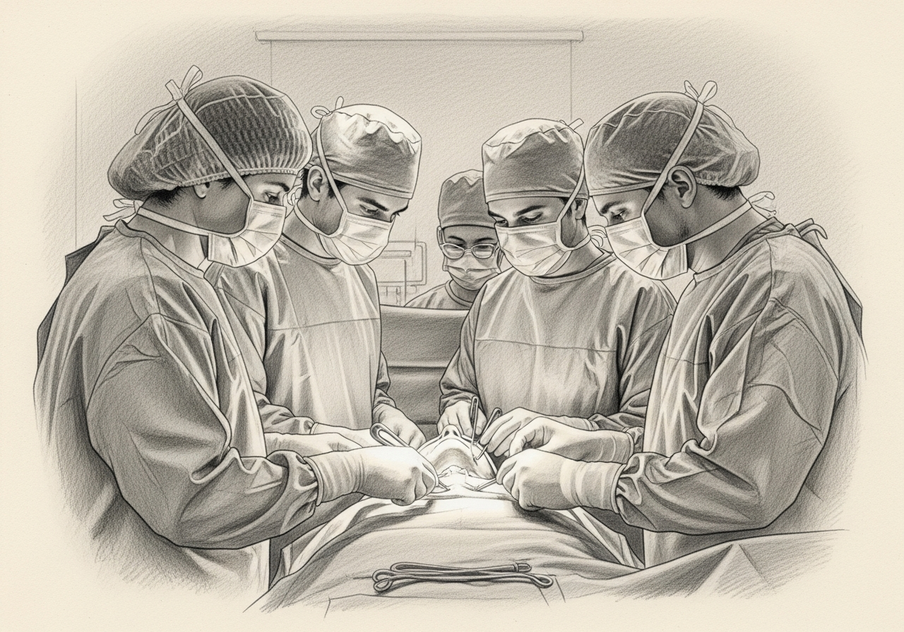
The operation is performed under general anesthesia through an incision in the upper neck below the jaw, placed in a natural skin crease when possible. The surgeon removes the gland while identifying and protecting the nearby nerves that control the lower lip and move the tongue and the nerve that supplies sensation and taste to part of the tongue.
When a stone sits within the gland or duct, the gland and stone are removed together. For tumors, the gland is removed intact for accurate pathology.
Recovery and aftercare
Many patients go home the same day or after an overnight stay. A small drain may be placed and removed within a day or two.
Temporary swelling, numbness, and a firm feeling along the incision are common, and some patients notice temporary changes in lip movement or tongue sensation that usually improve. Most patients resume light activity within one to two weeks with standard wound care.

Risks and alternatives
Risks may include temporary or, less commonly, lasting weakness of the lower lip, numbness or altered taste on part of the tongue, reduced tongue movement, bleeding, infection, salivary collection, and scarring. Risk depends on the size and nature of the gland problem.
Alternatives depend on the diagnosis and may include conservative care for infection, hydration and massage for mild stone disease, sialendoscopy for selected stones, or observation for some benign-appearing findings.
Results and follow-up
For tumors, removal allows a definitive diagnosis, and pathology guides any further treatment. For stones and chronic infection, removing the gland typically relieves recurrent swelling and infection.
Follow-up includes wound and nerve checks, scar care, and surveillance when a tumor was cancerous. Most people have no noticeable change in saliva because the remaining glands continue to function.
Clinical perspective
Our head and neck surgeons weigh whether the gland truly needs to be removed and how to protect the nerves that run beside it. Factors that make excision more appropriate include a tumor, recurrent infection, or chronic obstruction, and stones that cannot be cleared while preserving the gland. A stone that can be reached through the mouth or duct may instead favor a gland-preserving approach first.
The marginal mandibular, lingual, and hypoglossal nerves sit close to the gland, so the operation is planned to identify and protect them, and a tumor that involves these structures can change the recommendation. Candidacy and the right extent of surgery require individualized specialist review.
What to bring to your consultation
Gathering the right records helps a focused visit move quickly.
- Prior imaging and reports
- Pathology or biopsy results when applicable
- Recent laboratory results
- Treatment notes and a current medication list
- A clear note about the decision you want help with
A focused review covers the likely diagnosis, the realistic options, the trade-offs, and the steps needed before treatment. For urgent symptoms, follow the guidance above rather than using the routine form.
Medical review
This page is a patient-education resource reviewed by the responsible Norelle ENT clinician before publication. It does not replace an in-person evaluation. If symptoms are severe or rapidly worsening, seek immediate medical care.
Specialists who perform submandibular gland excision
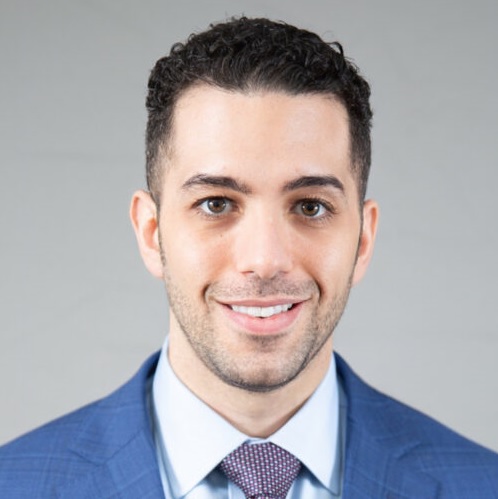
Dr. Moustafa Mourad
MD, FACS
Double Board-Certified Head & Neck and Facial Plastic & Reconstructive Surgeon
Dr. Moustafa Mourad is a double board-certified head and neck and facial plastic and reconstructive surgeon who cares for the full range of cosmetic and complex conditions affecting the face, head, and neck.
- Facial plastic and reconstructive surgery
- Head and neck cancer surgery
- Microvascular free-flap reconstruction
- Facial trauma and reconstruction
Also caring for this area


Not sure who to see? Our patient coordination team can help match you with the right specialist.
(212) 444-8006Frequently Asked Questions
It is a salivary gland located beneath the jaw on each side of the neck. It can develop tumors, stones, or chronic infection that may require removal.
Common reasons include a tumor, stones that repeatedly block the gland and cause swelling and infection, and chronic inflammation that does not respond to other care.
Nerves controlling the lower lip and tongue and providing tongue sensation run near the gland. Surgeons protect them, but temporary or, less commonly, lasting effects can occur.
It is placed in the upper neck below the jaw, within a natural skin crease when possible, to help it heal less noticeably.
Most people notice no meaningful change because the other salivary glands continue to produce saliva.
Some stones can be managed with hydration, massage, or sialendoscopy. Gland removal is considered when stones recur or cannot be reached, or when there is chronic damage.
For a gland mass, imaging and often a fine-needle biopsy are performed before surgery to help plan the operation.
Many patients resume light activity within one to two weeks, with swelling and numbness improving over the following weeks.
Some stones in the front or middle of the duct can be removed transorally. Deeper stones and scarred glands need individualized planning.
It provides general sensation to the front of the tongue. Injury can cause numbness or altered sensation.
It controls tongue movement. Weakness can affect speech and swallowing.
Clinical References
These independent resources from medical and professional organizations offer further reading. They are provided for general education and do not replace a consultation with a clinician.
Related Conditions

Related Procedures

Request a consultation about submandibular gland excision
Schedule a consultation with our team to discuss whether this procedure is the right option for you.