
Overview
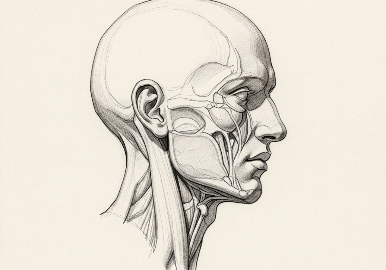
Salivary gland disease refers to a range of problems affecting the glands that produce saliva, including the parotid glands in front of the ears, the submandibular glands under the jaw, and the smaller sublingual and minor glands. Swelling can come from a stone or duct narrowing that blocks the flow of saliva, an infection, autoimmune inflammation, medication-related dryness, or a tumor, and the goal of evaluation is to tell these apart.
The timing of symptoms often provides the first clue. Swelling and pain that worsen around meals point toward obstruction, a painful and feverish gland suggests infection, and a persistent, painless lump raises concern for a tumor. Some conditions come and go, while others cause steady swelling or a growing mass that needs evaluation.
Norelle ENT evaluates salivary gland disease with examination, imaging, and tissue sampling when a tumor is suspected, and coordinates with radiology and pathology. Treatment ranges from conservative measures and gland-preserving duct procedures for stones and infections to surgery for persistent problems or tumors, with care to protect nearby nerves.
How we approach the decision
Salivary gland swelling has many possible causes, and the first task is to sort obstruction from infection, inflammation, autoimmune disease, and tumors. The decisions this care is meant to help you understand are:
- Is the problem obstructive, inflammatory, infectious, autoimmune, medication-related, or neoplastic?
- Can the diagnosis be made with ultrasound, CT or MRI, or needle biopsy without unnecessary gland removal?
- Is a gland-preserving duct procedure available and appropriate, or is excision the safer choice?
Living with salivary gland disease? The next step is a quiet, unhurried conversation.
What happens next
Evaluation usually follows a clear sequence:
- A history focused on meal-related swelling, pain, dry mouth, infections, medications, and autoimmune symptoms.
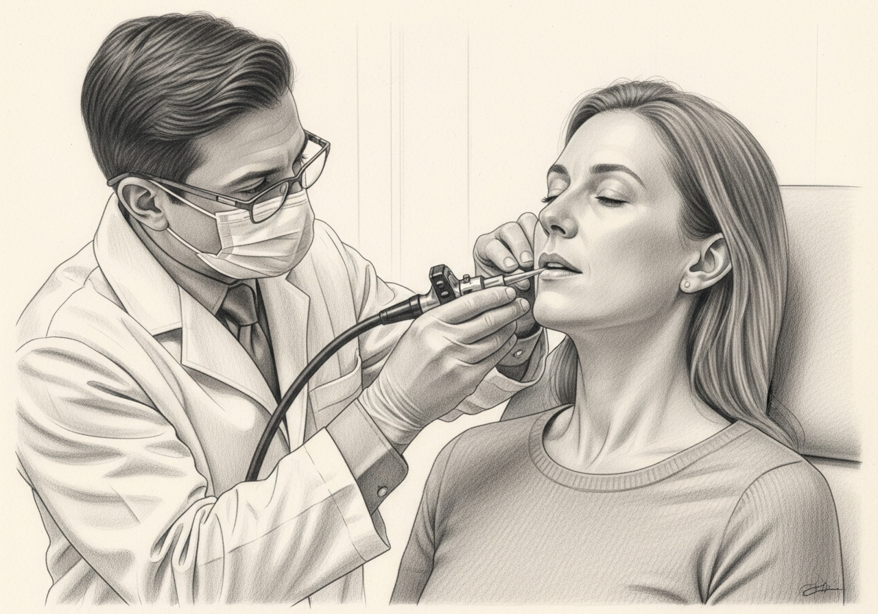
- Examination of the gland, duct opening, oral cavity, facial nerve, and neck.
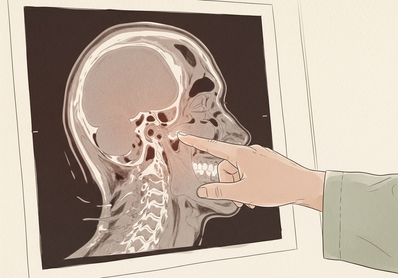
- Ultrasound or cross-sectional imaging based on the suspected cause.
- Fine needle aspiration or core biopsy for a persistent mass when appropriate, which may point toward a parotid tumor or salivary gland cancer.
- Treatment matched to the cause: hydration and duct care, medication, antibiotics when infection is present, stone or duct procedures, observation, or surgery. Gland-preserving and excisional options range from salivary gland surgery to parotidectomy or submandibular gland excision depending on which gland is involved.
Symptoms and warning signs
Common symptoms include swelling and pain in a gland, often worse around meals, and a persistent lump. Stones tend to cause intermittent swelling, while infections cause pain, redness, and sometimes fever.
A persistent or growing lump, especially when painless, should be evaluated to rule out a tumor. Facial weakness near the parotid gland or a hard, fixed mass deserves prompt attention.
Causes and risk factors
Causes of salivary gland disease include:
- Stones that block the flow of saliva
- Bacterial or viral infections
- Inflammatory and autoimmune conditions
- Dehydration and reduced saliva flow
- Benign and, less often, malignant tumors
Risk factors a clinician may ask about include dehydration, medications that reduce saliva, prior radiation, and autoimmune conditions.
How it is diagnosed
Evaluation begins with examination of the glands and ducts. Imaging and sampling are added based on the suspected cause:
- Ultrasound to look for stones, inflammation, or a mass
- CT or MRI for deeper assessment when needed
- Fine needle aspiration when a tumor is suspected
- Blood tests when an autoimmune cause is considered
The aim is to identify whether the problem is obstructive, infectious, inflammatory, or a tumor.
Treatment options
Treatment depends on the cause. Stones and mild infections often respond to hydration, warm compresses, gland massage, and antibiotics when needed. Some stones can be addressed with sialendoscopy, a minimally invasive technique that uses a tiny scope to access the duct.
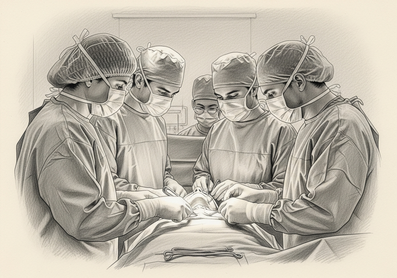
Inflammatory and autoimmune causes are treated medically. Persistent problems and tumors may require surgical removal of the gland, performed with care to protect nearby nerves, including the facial nerve near the parotid gland.
Salivary gland tumors
Tumors can develop in any salivary gland, and the likelihood of a tumor being benign or cancerous varies by gland and by the type of cells involved. Many tumors of the large glands are benign, but a portion are cancerous, and tumors of the smaller glands carry a different mix of possibilities.
A persistent, painless lump is the most common sign of a tumor. Fine needle aspiration and imaging help suggest the type, but the final diagnosis is confirmed on the tissue removed at surgery, and the plan depends on whether the tumor is benign or malignant and on its location.
Facial nerve considerations
The facial nerve runs through the parotid gland, and other important nerves lie near the submandibular and other glands. Surgery on these glands is planned to identify and preserve these nerves whenever possible, and nerve monitoring may be used.
When a cancer involves a nerve, the surgeon discusses the implications, which may include repair or grafting. Temporary changes in facial movement, sensation, or taste can occur even when nerves are preserved, and these are reviewed before surgery.
Recovery after surgery
Recovery after salivary gland surgery depends on which gland is treated and the extent of the operation. It may include a short period of wound care, a drain, and temporary swelling or numbness near the incision.
The care team reviews activity, diet, and follow-up, including monitoring of nerve function. When surgery is performed for cancer, follow-up includes a schedule of examinations and imaging based on the diagnosis, and additional treatment such as radiation is discussed when it is recommended.

When to seek urgent care
Use these categories to guide timing:
- Emergency, meaning call 911 or go to the nearest emergency department: rapidly increasing painful swelling with fever, difficulty breathing, difficulty swallowing, or spreading redness with signs of a serious infection.
- Same-day or urgent evaluation: a painful, febrile gland or quickly worsening swelling, dehydration, or new facial weakness.
- Routine specialist evaluation: recurrent gland swelling, swelling that worsens around meals, or a persistent, painless lump that should be evaluated to rule out a tumor.
The online consultation form is for routine scheduling and is not an emergency service.
Clinical perspective
Our head and neck surgeons often start from the symptom pattern. Swelling that comes with meals suggests obstruction from a stone or duct narrowing. A painful, febrile gland suggests infection. A painless, persistent lump raises concern for a tumor and usually needs imaging and tissue diagnosis. Dry mouth affecting several glands can point to an autoimmune cause such as Sjogren disease. Facial weakness with a parotid mass is a feature that calls for prompt, expedited evaluation.
Factors that generally favor gland-preserving treatment include stones or duct narrowing that can be reached with a duct procedure, and infection or inflammation that responds to medical care. Factors that call for caution, and may favor surgery, include a persistent firm mass, a tumor, or recurrent disease that does not settle. Antibiotics are used when bacterial infection is suspected, not for every episode of swelling.
What commonly changes the recommendation is the timing of symptoms, ultrasound and biopsy findings, and whether gland-preserving techniques are suitable. Candidacy is determined individually after specialist review rather than from a general rule.
What to bring to your consultation
Bringing the right records makes a consultation more efficient. Helpful items include:
- Prior imaging and the written reports
- Pathology or biopsy results when available
- Notes on the timing of symptoms, including whether swelling worsens with meals
- Treatment notes from any prior care, including prior infections or procedures
- A current medication list
- The specific decision you want the consultation to answer
Request a consultation for a focused review of the diagnosis, the available options, the likely tradeoffs, and the steps needed before treatment. For urgent symptoms, follow the guidance above rather than using the routine form.
Medical review
This page is a patient-education resource reviewed by the responsible Norelle ENT clinician before publication. It does not replace an in-person evaluation. If symptoms are severe or rapidly worsening, seek immediate medical care.
Specialists who treat salivary gland disease
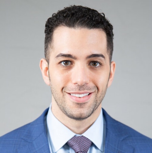
Dr. Moustafa Mourad
MD, FACS
Double Board-Certified Head & Neck and Facial Plastic & Reconstructive Surgeon
Dr. Moustafa Mourad is a double board-certified head and neck and facial plastic and reconstructive surgeon who cares for the full range of cosmetic and complex conditions affecting the face, head, and neck.
- Facial plastic and reconstructive surgery
- Head and neck cancer surgery
- Microvascular free-flap reconstruction
- Facial trauma and reconstruction
Also caring for this area

Not sure who to see? Our patient coordination team can help match you with the right specialist.
(212) 444-8006Norelle Academic Work
Research by Our Physicians
Selected scholarly work related to this topic.

Commentary on “Incisions for Parotidectomy: A Narrative Review”
Annals of Plastic Surgery
Cost-Effectiveness Analysis of Transfacial Gland-Preserving Removal of Parotid Sialoliths
The Laryngoscope
Pull-Through Sialodochoplasty for Stensen’s Megaduct
The Laryngoscope
Increased Local Recurrence in Advanced Parotid Malignancy Treated with Mastoidectomy without Lateral Temporal Bone Resection
Oral and Maxillofacial Surgery
Citation details link to the original PubMed, DOI, journal, or publisher record.
Explore all Norelle research on this topicFrequently Asked Questions
Salivary gland disease includes stones that block saliva, infections, inflammation, autoimmune conditions, and benign or malignant tumors. Evaluation identifies which is present so treatment can be directed accurately.
Eating stimulates saliva production. If a stone or narrowing blocks the duct, saliva backs up and the gland swells and becomes painful around meals, then often settles afterward.
Evaluation includes an examination, imaging such as ultrasound, CT, or MRI, and fine needle aspiration when a tumor is suspected. Blood tests are added when an autoimmune cause is considered.
Sialendoscopy is a minimally invasive technique that uses a very small scope to access the salivary duct, allowing some stones and narrowings to be treated while preserving the gland.
No. Many stones pass or are managed with hydration, massage, and warm compresses. Larger or persistent stones may need sialendoscopy or, in some cases, surgery.
A persistent or growing lump, especially when painless, should be evaluated to rule out a tumor. A hard, fixed mass or facial weakness near the parotid gland deserves prompt attention.
Often, yes. Stones, infections, and inflammation are frequently managed without removing the gland. Surgery is reserved for persistent problems or tumors, and it is planned to protect nearby nerves.
Many tumors of the large salivary glands are benign, but a portion are cancerous, and the likelihood varies by gland and tumor type. Evaluation with imaging and a needle biopsy helps clarify the diagnosis.
Surgery near the parotid gland is planned to identify and preserve the facial nerve, and nerve monitoring may be used. Temporary weakness can occur even when the nerve is preserved, and the surgeon reviews these possibilities beforehand.
No. Radiation is recommended in selected cases based on the tumor type, grade, and surgical findings. Whether it is advised is discussed after the diagnosis is confirmed.
A persistent or growing lump, especially when painless or firm, is usually assessed with imaging and fine needle aspiration to help clarify whether it is benign or malignant before treatment is planned.
Yes. Autoimmune conditions such as Sjogren disease can cause dry mouth and swelling in several glands, and blood tests are added when an autoimmune cause is considered.
Antibiotics are used when a bacterial infection is suspected, not for every episode of swelling. Obstruction, inflammation, and tumors are treated differently, so the cause is identified first.
Clinical References
These independent resources from medical and professional organizations offer further reading. They are provided for general education and do not replace a consultation with a clinician.
Related Procedures

Related Conditions

Request a consultation for salivary gland disease
Schedule an evaluation with our team to review your symptoms and the appropriate next steps.