

About the Procedure
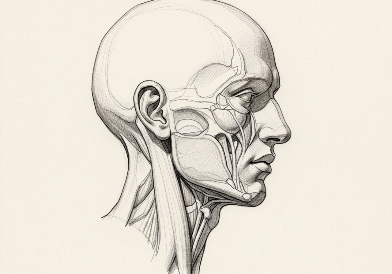
A free flap, also called free tissue transfer, is an advanced reconstructive technique used to repair complex defects of the head and neck after tumor removal, severe trauma, or radiation injury. Tissue, which may include skin, fat, muscle, or bone, is taken from a donor site such as the forearm, thigh, or lower leg, along with its blood vessels.
What distinguishes a free flap from a local flap is that the tissue is completely detached and then reconnected to blood vessels near the defect using microsurgery. This allows healthy, well-vascularized tissue to be moved to areas that cannot be reconstructed with nearby tissue alone.
At Norelle Health, free flap reconstruction is planned as part of a coordinated effort to restore both form and function, such as the ability to speak, swallow, or support facial structure. The donor site and flap type are chosen to match the needs of the defect and the patient's overall health.
How we approach the decision
Free flap planning focuses on three decisions:
- What tissue characteristics are needed: thin lining, bulk, bone length, a skin paddle, nerve, or multiple components?
- Which donor site provides the closest match with the least functional cost?
- How will the flap be monitored, and what is the rescue pathway if blood flow becomes compromised?
These questions connect the defect to the reconstruction. Free flap reconstruction is one part of broader head and neck reconstruction and is often planned alongside oral cancer surgery or other head and neck cancer surgery.
Considering free flaps? The next step is a quiet, unhurried conversation.
What happens next
The pathway from planning to recovery generally follows these steps:
- Map the defect and choose candidate donor sites.
- Assess vessels, prior treatment, vascular disease, mobility, hand or leg dominance, and rehabilitation needs.
- Perform tumor resection and flap harvest with coordinated teams when appropriate.
- Reconnect the artery and vein and monitor perfusion intensively.
- Advance wound, airway, feeding, mobility, donor-site, speech, and swallowing care, supported by structured head and neck surgery recovery and nutrition after surgery.
When to seek urgent care
A sudden change in flap color, warmth, swelling, bleeding pattern, Doppler signal, or wound appearance is time-sensitive. In the hospital, staff monitor the flap closely; after discharge, report the specific warning signs your team provides.
- Emergency (call 911 or go to the nearest emergency department): significant bleeding, breathing difficulty, or rapidly increasing swelling.
- Same-day contact with the surgical team: a change in flap color or temperature, increasing pain, or wound drainage after discharge.
- Routine: general questions about healing or the donor site can be handled through a scheduled visit.
The online consultation form is not monitored for emergencies and should never be used for urgent symptoms.
Clinical perspective
Our head and neck surgeons select a donor site to match the tissue the defect requires while limiting the functional cost of harvest. Commonly used options include:
- Radial forearm: thin, pliable tissue useful for lining and soft-tissue defects; it leaves a forearm scar and is assessed for hand circulation before use.
- Anterolateral thigh: versatile skin and soft tissue with adjustable bulk and a generally well-tolerated donor site.
- Fibula: a long segment of vascularized bone with a skin paddle for jaw reconstruction, with leg circulation and mobility evaluated beforehand.
- Scapular system: combined bone and soft tissue useful for complex composite defects.
Factors that make a flap a stronger choice include a good tissue and vessel match and an acceptable donor-site effect; factors that require caution include prior radiation, vascular disease, prior surgery, or limited recipient vessels. No single donor site suits every patient, and candidacy requires individualized specialist review.
What to bring to your consultation
A focused review is easier when you bring the records most relevant to microvascular free flap reconstruction:
- Prior imaging and radiology reports.
- Pathology or biopsy results when available.
- Recent laboratory results.
- Prior treatment notes, including any radiation.
- A current medication list.
- The specific decision you want the consultation to answer.
Request a consultation for a focused review of the diagnosis, the available options, the likely tradeoffs, and the steps needed before treatment. For urgent symptoms, follow the emergency guidance above rather than using the routine form.
Who may be a candidate
A free flap may be considered when:
- A defect is too large or complex for local reconstruction
- Tissue is needed after cancer removal, trauma, or radiation
- Healthy donor and recipient blood vessels are available
- Overall health supports a longer operation
The flap type and donor site are chosen to match the tissue needs of the defect.
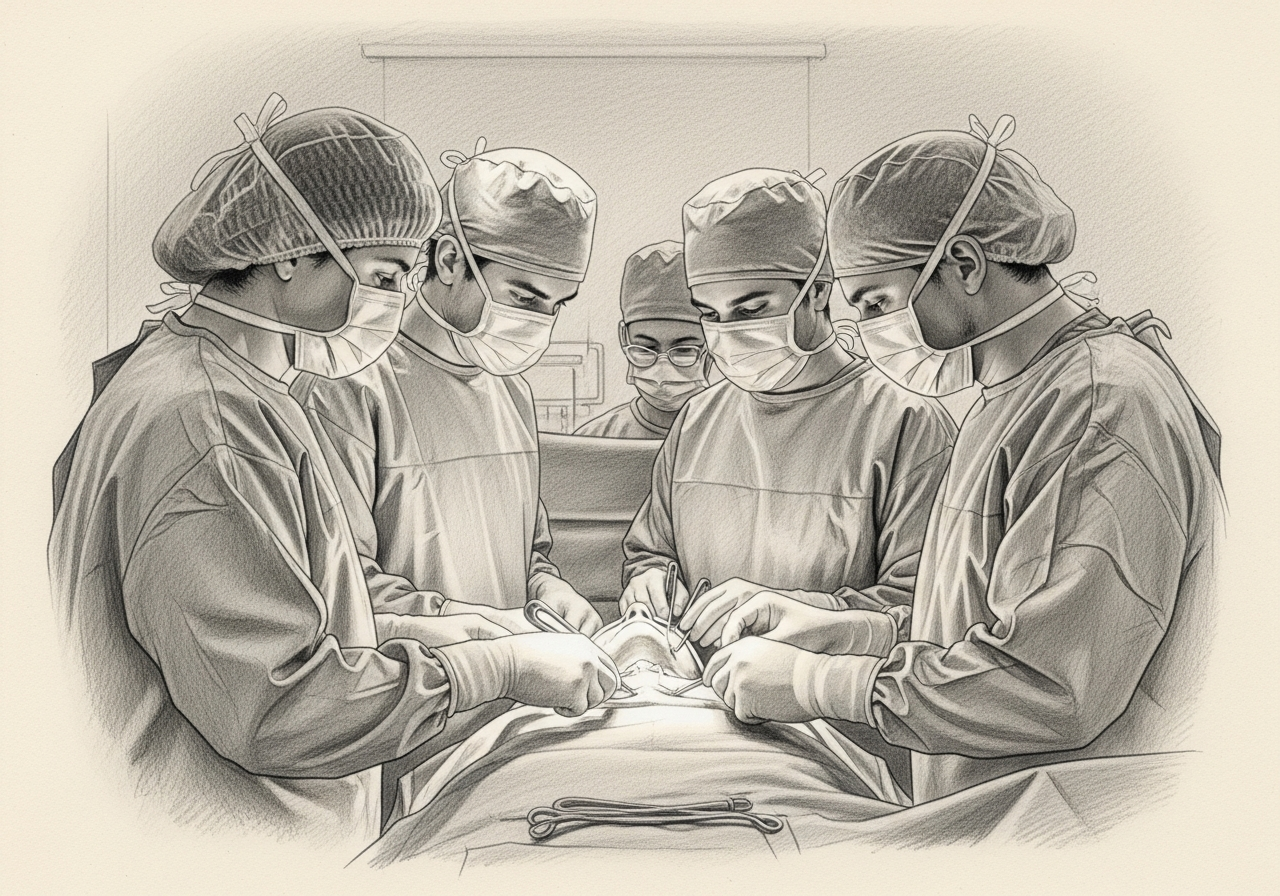
How it is performed
Under general anesthesia, tissue, which may include skin, fat, muscle, or bone, is raised from a donor site along with its supplying blood vessels. The tissue is shaped to the defect, and its blood vessels are reconnected to vessels near the reconstruction site using microsurgery.
The donor site is then closed, sometimes with a skin graft. The operation is detailed and typically takes several hours.
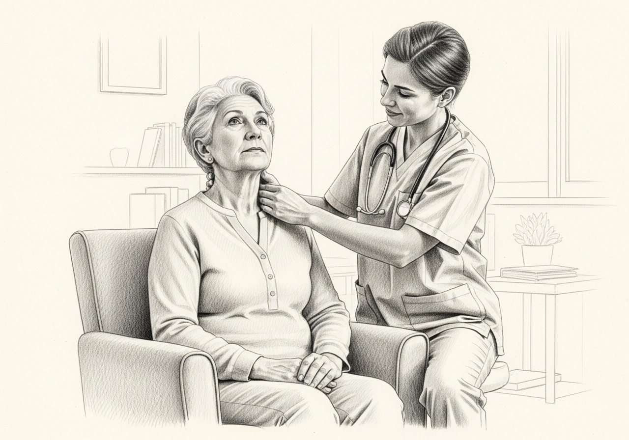
Recovery and aftercare
After surgery, the flap's blood supply is monitored closely, often in a hospital setting, during the early days when complications are most likely. Healing continues at both the reconstruction and donor sites over the following weeks.
Depending on what was reconstructed, therapy may help restore speech, swallowing, or movement, and function can keep improving over months.
Risks and alternatives
Possible risks include partial or complete flap loss if the blood supply fails, bleeding, infection, donor-site healing problems, scarring, and the need for additional procedures. Close monitoring helps detect and address problems early.
Alternatives may include local or regional flaps, skin grafts, or, in some cases, simpler closure, depending on the defect. The best option is determined by the reconstruction team.
Results and follow-up
Free flap reconstruction is intended to restore structure and function and to support healing in areas affected by cancer surgery, trauma, or radiation. Results develop as healing progresses.
Follow-up monitors healing, function, and, when relevant, surveillance for the underlying condition, with revisions sometimes performed to refine the result.
Medical review
This page is a patient-education resource reviewed by the responsible Norelle Health clinician before publication. It does not replace an in-person evaluation. If symptoms are severe or rapidly worsening, seek immediate medical care.
Specialists who perform free flaps
Dr. Moustafa Mourad
MD, FACS
Double Board-Certified Head & Neck and Facial Plastic & Reconstructive Surgeon
Dr. Moustafa Mourad is a double board-certified head and neck and facial plastic and reconstructive surgeon who cares for the full range of cosmetic and complex conditions affecting the face, head, and neck.
- Facial plastic and reconstructive surgery
- Head and neck cancer surgery
- Microvascular free-flap reconstruction
- Facial trauma and reconstruction
Also caring for this area

Not sure who to see? Our patient coordination team can help match you with the right specialist.
(212) 444-8006Frequently Asked Questions
A free flap, or free tissue transfer, moves living tissue with its own blood vessels from a donor site to repair a complex defect, reconnecting the vessels under a microscope.
A local flap uses nearby tissue that stays attached to its blood supply, while a free flap is fully detached and its vessels are reconnected with microsurgery, allowing tissue to be moved from a distant donor site.
Free flaps are used for large or complex head and neck defects that cannot be repaired with local tissue, often after cancer surgery, severe trauma, or radiation injury.
Donor sites vary by need and may include the forearm, thigh, or lower leg, and the flap can include skin, fat, muscle, or bone.
Recovery usually begins with a hospital stay and close monitoring of the flap, followed by healing over weeks. Functional recovery, such as speech or swallowing, can continue to improve over months.
If the blood supply to the flap fails, part or all of the flap can be lost, sometimes requiring additional surgery. Close early monitoring is used to detect and address problems quickly.
Yes. The donor site is closed and leaves a scar, and it is sometimes covered with a skin graft. The team selects a donor site to balance the reconstruction needs and donor-site effects.
Depending on the defect, alternatives may include local or regional flaps, skin grafts, or simpler closure. The reconstruction team recommends the most suitable approach.
A free flap is completely detached and reconnected to neck vessels microsurgically. A regional flap remains attached to its original blood supply and is rotated into the defect.
Monitoring is most frequent early after surgery, when vascular problems are most likely and timely re-exploration can matter. Exact protocols vary.
It can provide a long segment of vascularized bone with skin and soft tissue, while generally preserving useful leg function in appropriately selected patients.
It may include vascular examination or imaging, assessment of mobility and medical conditions, and evaluation of the specific functional cost.
Clinical References
These independent resources from medical and professional organizations offer further reading. They are provided for general education and do not replace a consultation with a clinician.
Related Conditions

Related Procedures

Request a consultation about free flaps
Schedule a consultation with our team to discuss whether this procedure is the right option for you.