
About the Procedure
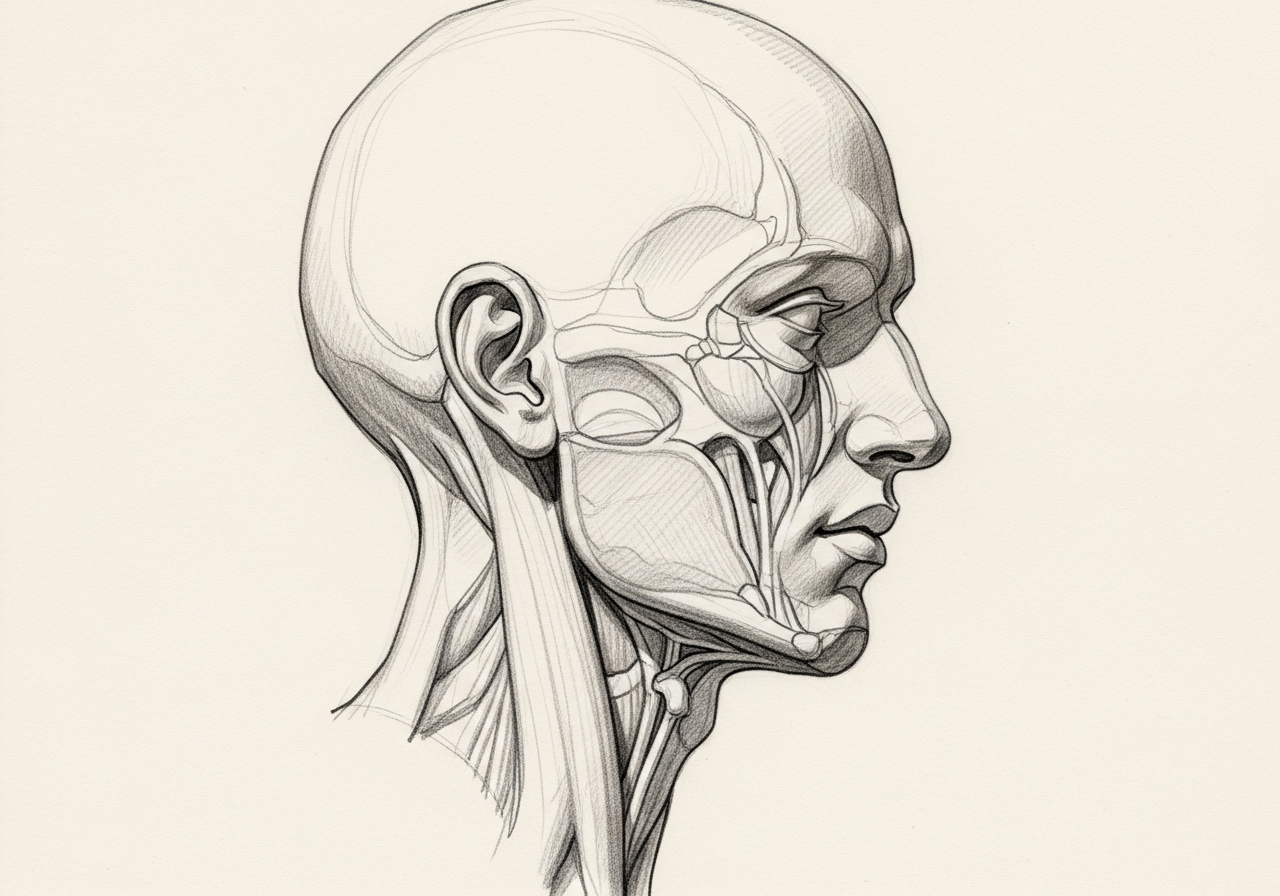
Cranioplasty is surgery to rebuild an opening or weakened area in the skull vault, also called the calvarium. The defect may remain after a decompressive craniectomy, trauma, tumor removal, infection, or a prior operation. Unlike endoscopic skull-base reconstruction, which seals the barrier between the brain and the nose or sinuses, cranioplasty restores the protective outer framework around the brain.
The reconstruction may use the patient's preserved bone when it remains suitable or an implant made from materials such as titanium or a patient-specific synthetic material. The choice depends on the defect's size and location, the condition of the scalp and surrounding tissue, prior infection or radiation, previous operations, imaging, and the patient's overall health.
Planning is multidisciplinary. Neurosurgery addresses the brain, its coverings, and the safety of replacing the missing skull, while reconstructive surgeons may help plan scalp coverage, contour, and healing in complex or previously operated areas. The goal is a durable repair that protects the brain, restores contour, and avoids adding more risk than the expected benefit justifies.
How we approach the decision
Cranioplasty is planned around the reason for the skull defect and the condition of the brain, scalp, and remaining bone. A consultation should clarify several decisions.
- Is the brain swelling controlled and is the patient medically and neurologically ready for reconstruction?
- Is there active or recent infection, poor scalp coverage, prior radiation, or another factor that changes timing or material choice?
- Can the patient's own bone be used safely, or would a patient-specific implant offer a more reliable fit?
- Will the scalp close without excessive tension, or is additional reconstructive tissue needed?
This is different from endoscopic skull-base reconstruction, which repairs a leak or barrier defect between the cranial cavity and the nose or sinuses.
Considering cranioplasty? The next step is a quiet, unhurried conversation.
What happens next
Evaluation and treatment usually follow a coordinated sequence.
- Review the original operation, neurologic course, wound history, infection history, and current symptoms.
- Examine the scalp and defect and review thin-cut CT imaging to define the bone opening and plan the repair.
- Confirm that swelling and infection are controlled and compare autologous bone with available implant options.
- Coordinate neurosurgical exposure and protection of the brain with any scalp or soft-tissue reconstruction needed for durable coverage.
- Monitor neurologic status, the wound, and the reconstruction after surgery, then advance activity according to healing.
Why cranioplasty may be recommended
A skull defect can leave the brain less protected and may create a visible or palpable contour change. Some patients also have headaches, discomfort, or neurologic symptoms after a large decompressive craniectomy, although no website can predict whether reconstruction will improve a particular symptom.
Cranioplasty may restore the skull's protective structure and contour and can be an important stage of recovery after the condition that required removal of bone has stabilized. The expected benefits must be weighed against the risks of another operation.
Choosing bone or an implant
The patient's preserved bone flap may be an option when it is available, structurally sound, and free of infection. In other situations, surgeons may use titanium or a synthetic implant shaped from CT data to match the defect.
No material is best for every patient. Autologous bone can resorb or become infected; implants can also become infected, loosen, fracture, or require removal. Defect size, age, prior contamination, scalp quality, cost and availability, and the need for a custom contour all affect the choice.
Timing and infection control
Cranioplasty is generally delayed until the patient is stable, brain swelling has settled, and the wound is ready for another operation. The exact timing varies with the reason for the original craniectomy, neurologic recovery, infection risk, and the need for other treatment.
Suspected infection, drainage, exposed bone or hardware, or unhealthy scalp tissue may require treatment before a permanent reconstruction. In complex cases, removal of infected material, antibiotics, wound reconstruction, and later cranioplasty may be staged rather than performed at one operation.
Scalp and soft-tissue reconstruction
A durable cranioplasty needs healthy, well-vascularized tissue over the bone or implant. Prior surgery, trauma, infection, and radiation can leave the scalp thin, scarred, or tight.
When simple closure would place too much tension on the wound, reconstructive options may include rearranging nearby scalp, tissue expansion, a regional flap, or free tissue transfer. The purpose is reliable coverage of the reconstruction, not simply closing the skin.
Risks and alternatives
Risks can include bleeding, infection, wound breakdown, fluid collection, seizure, injury to the brain, stroke, blood clot, contour irregularity, bone resorption, implant exposure or failure, and the need for another operation. Individual risk depends heavily on the original neurologic condition, prior infection, scalp quality, and medical health.
Alternatives may include delaying reconstruction while the brain or wound recovers, treating infection first, using protective headgear, revising the scalp before placing an implant, or not proceeding when the operative risk outweighs the expected benefit. The neurosurgical and reconstructive teams should explain the trade-offs for the specific defect.
Recovery and follow-up
After surgery, the team monitors neurologic function, the incision, swelling, and any fluid collection. Imaging may be obtained when symptoms or the operative plan call for it. Temporary activity restrictions protect the wound and reconstruction while healing begins.
Recovery also reflects the condition that led to the skull defect. Some patients continue neurologic rehabilitation independent of the cranioplasty itself. Contact sports and activities with head-impact risk require explicit clearance, and a helmet should be used or discontinued only as directed by the treating team.
When to seek urgent care
After cranioplasty, call the surgical team promptly for increasing redness, drainage, wound opening, fever, worsening swelling, or escalating pain. Seek emergency care for a new seizure, new weakness or numbness, confusion, loss of consciousness, severe or rapidly worsening headache, repeated vomiting, major bleeding, or another sudden neurologic change.
These symptoms cannot be assessed through an online form. The treating team's discharge instructions take priority.
What to bring to your consultation
Where possible, bring or securely transfer:
- CT and MRI images and reports, not only the written reports
- The operative report from the craniectomy or prior reconstruction
- Culture results and antibiotic records if infection occurred
- Implant information and prior pathology reports when applicable
- Recent neurosurgery, rehabilitation, and wound-care notes
- A current medication list, especially blood thinners and seizure medicines
- The specific decision you want help evaluating
Medical review
This physician-reviewed page is a patient-education resource and does not replace an examination by the neurosurgical and reconstructive teams responsible for the defect. New neurologic symptoms, a seizure, wound drainage, rapidly increasing swelling, or severe worsening headache after skull surgery require urgent assessment.
Specialists who perform cranioplasty
Dr. Moustafa Mourad
MD, FACS
Double Board-Certified Head & Neck and Facial Plastic & Reconstructive Surgeon
Dr. Moustafa Mourad is a double board-certified head and neck and facial plastic and reconstructive surgeon who cares for the full range of cosmetic and complex conditions affecting the face, head, and neck.
- Facial plastic and reconstructive surgery
- Head and neck cancer surgery
- Microvascular free-flap reconstruction
- Facial trauma and reconstruction
Also caring for this area


Not sure who to see? Our patient coordination team can help match you with the right specialist.
(212) 444-8006Norelle Academic Work
Research by Our Physicians
Selected scholarly work related to this topic.

Cranioplasty
Facial Plastic Surgery
Calvarial Reconstruction
Seminars in Plastic Surgery
Citation details link to the original PubMed, DOI, journal, or publisher record.
Explore all Norelle research on this topicFrequently Asked Questions
No. Cranioplasty rebuilds the skull vault around the brain. Skull-base reconstruction usually repairs a barrier defect between the cranial cavity and the nose, sinuses, ear, or other deep spaces, often to prevent a cerebrospinal fluid leak.
There is no single schedule for everyone. Timing depends on control of brain swelling and infection, wound healing, neurologic stability, medical fitness, and the reason the bone was removed. The treating neurosurgeon determines when the balance of risk and benefit is favorable.
No. Preserved bone may be suitable in some cases. Titanium or synthetic patient-specific implants may be preferred when bone is unavailable, contaminated, damaged, resorbed, or unlikely to provide a durable contour.
Some patients report improvement in selected symptoms after reconstruction of a large defect, but results are not predictable and depend on the underlying brain injury or disease. The primary goals are protection and restoration of the skull's structure and contour.
The team may need to improve the soft-tissue coverage before or during cranioplasty. Options range from local scalp rearrangement to tissue expansion or transfer of vascularized tissue, depending on the size and condition of the wound.
Yes. Infection is an important risk and may require antibiotics, drainage, or removal of the bone or implant. Prior infection, repeated surgery, wound problems, and poor tissue quality can increase risk.
Clinical References
These independent resources from medical and professional organizations offer further reading. They are provided for general education and do not replace a consultation with a clinician.
Related Procedures

1 of 3 · Head and Neck Reconstruction
Request a consultation about cranioplasty
Schedule a consultation with our team to discuss whether this procedure is the right option for you.